Orbital reconstruction, CAS: Intraoperative imaging
1. Introduction
When using computer assisted surgery (CAS) and intraoperative imaging the reduction (and fixation if necessary) is performed according to standard procedures described in the AO Surgery Reference. CAS should be considered as an adjunct to surgical treatment.
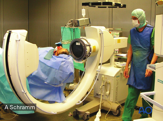
When treating orbital fractures CAS allows intraoperative visualization of the reconstruction using intraoperative imaging combined with image fusion of preoperative and intraoperative CT scans.
Intraoperative imaging assists in controlling the position of the implant during orbital reconstruction. Though it improves the surgical outcome and avoids the need for revision surgery, it also increases the duration of the operation and the radiation exposure.
In simple orbital floor fractures, radiopaque material for orbital floor reconstruction can easily be visualized by intraoperative imaging resulting in intraoperative or postoperative verification of implant placement.
With this technique, insufficient orbital reconstruction can be identified and corrected, eliminating the need for secondary procedures that may be necessary if only postoperative imaging is performed.
Computer assisted orbital reconstruction planning
Reconstruction of complex orbital defects may benefit from the preoperative virtual insertion of anatomically preformed orbital implants.
Some complex zygomatico-orbital defects may require patient-specific implants for alloplastic reconstruction of the orbit. This can be performed by transferring virtual preoperative planning into CAD-CAM tools to create patient-specific alloplastic implants or by pre-bending standard implants using rapid prototyping/stereolithographic models made from patients' anatomy or a virtually preplanned reconstruction.
2. Intraoperative assessment of reconstruction
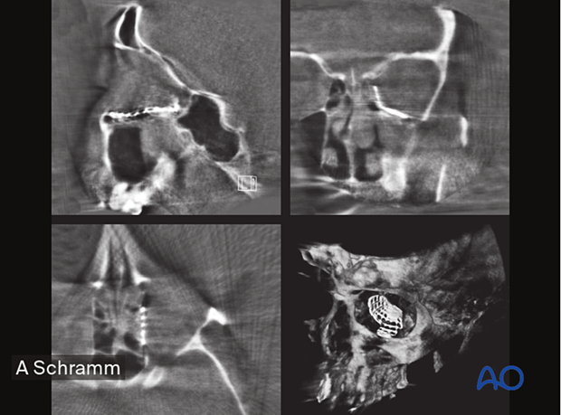
A CT scan is performed intraoperatively to verify that the orbit has been appropriately reconstructed.

The correct anatomic shape and position of the titanium mesh used for orbital wall reconstruction can be verified in the intraoperative CT scan. Due to the smaller radiation volumes used in intraoperative CT scans, the evaluation of symmetry is limited. Bigger scan volumes should be avoided due to higher radiation doses.


