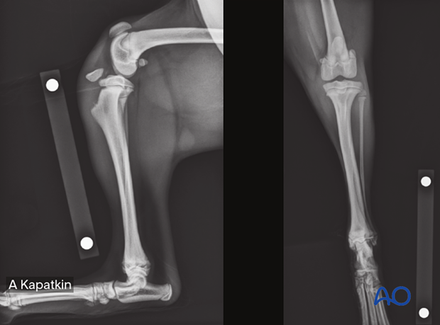
Release notes - Dog proximal tibia
1. First edition
Publication of module (July 21, 2025)
The module on this anatomical area has been authored by Kenneth Johnson (Australia). Amy Kapatkin (USA) acted as editor.
The dog proximal tibia module includes pages on:
- Fracture diagnosis
- Approaches to the proximal tibia
- Positioning
- Complications
- Wide selection of fracture fixation techniques tailored to the specific fracture types
Around 300 original illustrations accompany the step-by-step descriptions of the various fixation techniques and approaches.
To help identify the fractures, x-rays of most fracture types have been included in the diagnosis section, and the fixation techniques have been enriched with case examples with pre- and postoperative, and follow-up x-rays.
This module is exclusively for AO VET members only.
Go to the module entry page.

Table of contents
The dog proximal tibia material includes a detailed description of the following fracture types and fixation methods.
41-A1 Avulsion fracture:
- Nonoperative treatment
- K-wire fixation
- K-wire with tension band fixation
41-A2 Simple extraarticular fracture:
- Nonoperative treatment
- Bone plate fixation
- K-wire fixation
41-A3 Multifragmentary extraarticular fracture:
- Bridging plate fixation
41-B1 Lateral simple partial articular fracture:
- Lag screw and K-wire fixation
- Lag screw and buttress plate fixation
41-B2 Medial simple partial articular fracture:
- Lag screw and K-wire fixation
- Lag screw and buttress plate fixation
41-B3 Multifragmentary partial articular unicondylar fracture:
- Buttress plate and position screw or K-wire fixation
41-C1 Simple articular, simple metaphyseal fracture:
- Lag screw and neutralization plate fixation
41-C2 Simple articular, multifragmentary metaphyseal fracture:
- Salvage procedure
41-C3 Multifragmentary articular fracture:
- Salvage procedure
In addition, the material contains information about the following:
- Diagnosing each fracture type
- Indications for all treatments
- Dorsal recumbency positioning
- Craniomedial, craniolateral, and cranial approaches to the proximal tibia
- Basic techniques for lag screw fixation, plate preparation, and K-wire fixation


