

Lag screw and neutralization plate fixation
1. Indications
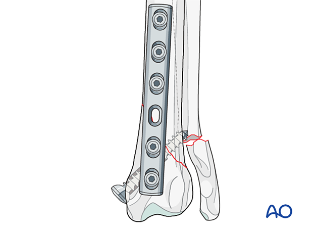
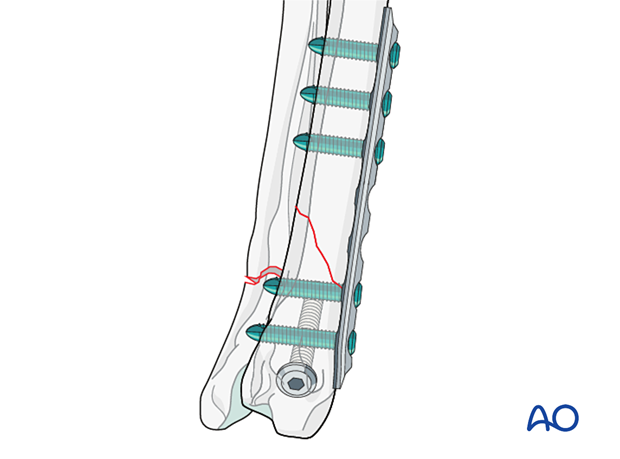
This technique is used to achieve interfragmentary compression for anatomically reconstructable long oblique or butterfly fractures.
With this technique, absolute stability is achieved. The lag screw provides primary fixation of the radius and applies interfragmentary compression to the fracture. The neutralization plate protects the primary fixation and offers axial stability.
2. Preparation and approach
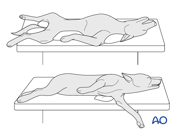
The patient can be in dorsal or lateral recumbency.
In lateral recumbency, the affected leg should be in contact with the table.
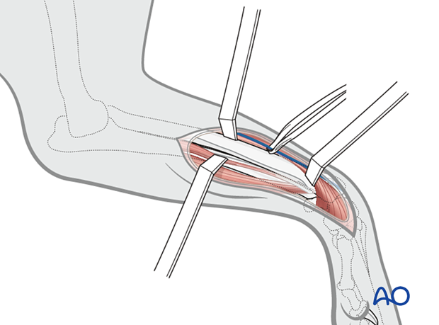
A craniomedial approach to the distal radius is performed.
3. Lag screw fixation
Reduction of the fracture line
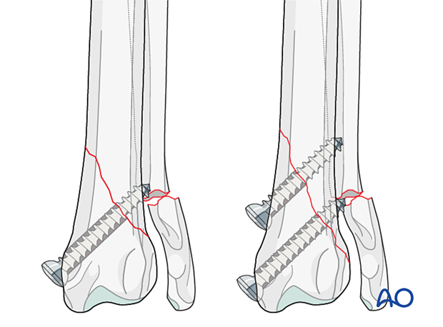
In an oblique fracture pattern, the bones are slid along the fracture line into anatomical reduction with the help of one or two pointed reduction forceps placed across the fracture line.

Fixation of the fracture
The oblique fracture is secured with a lag screw.
Read more about lag screw fixation.
If the fracture length allows for insertion of two screws, this is recommended.
Alternative: Lag screw through neutralization plate
If the plate is applied on the medial side, as an alternative to fixation with independent lag screws, the fracture can also be fixed with lag screws inserted through the neutralization plate.
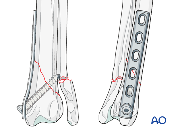
4. Neutralization plating
Plate selection
According to the size of the distal fragment, a straight plate or a T-plate should be used in order to have a minimum of two screws in the distal fragment and three screws in the proximal fragment.
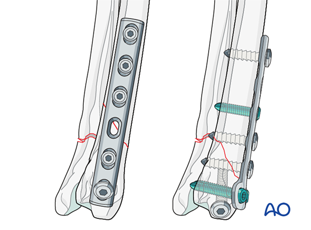
Plate contouring
The plate must be meticulously contoured to the bone to avoid fragment displacement and loss of reduction.
Read more about plate preparation.
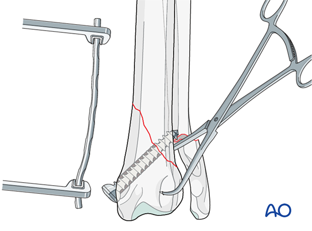
Plate application
If a straight plate is selected, it can be used on the cranial or medial side of the distal radius, depending on the fracture configuration.
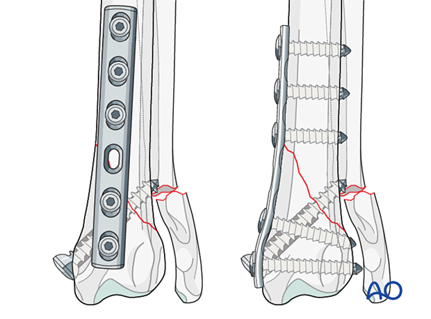
In more distal fractures or in toy breed dogs with limited bone stock, a T-plate may be required and should be positioned on the cranial side. See the corresponding treatment “T-plate fixation”.
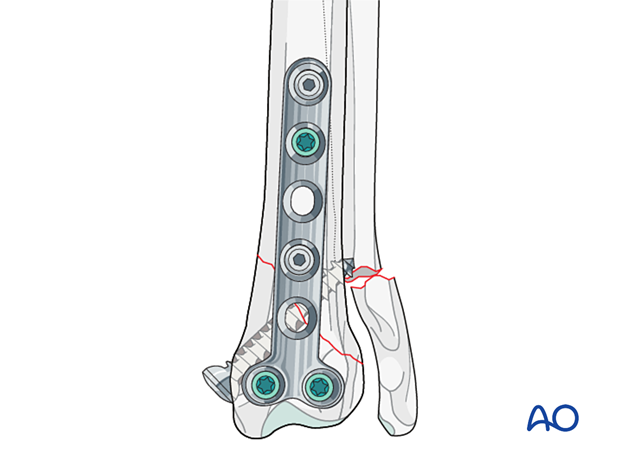
The contoured plate is applied to the distal radial surface and secured with plate-holding forceps.
If required, the alignment is corrected, and the plate-holding forceps are repositioned.
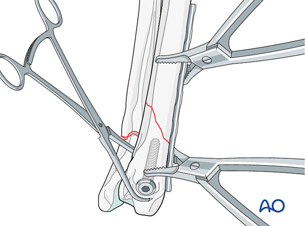
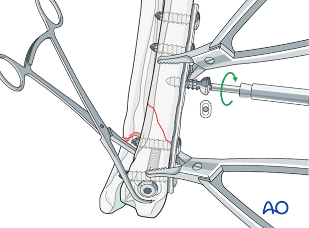
Fixation with a nonlocking plate
Screw insertion close or at the level of the fracture line should be avoided. All cortical screws are placed in neutral mode. Screw insertion order is not critical.
Fixation with a locking compression plate
A push-pull device can be inserted instead of plate-holding forceps in the proximal and distal fragment to temporarily stabilize the plate if a locking compression plate (LCP) is used.
With the temporary fixation in place, plate position and anatomical reduction are checked thoroughly and, if necessary, adjusted.
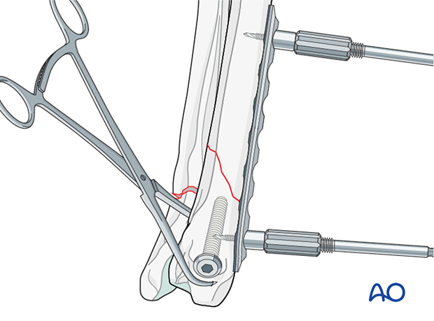
Locking plates lend themselves well to neutralization plating and do not require perfect contouring. Additionally, locking screws provide superior fixation to the bone. If an LCP is used, two to three locking screws per main fragment are needed.
5. Aftercare
Phase 1: 1–3 days after surgery
The aim is to reduce edema, inflammation, and pain.
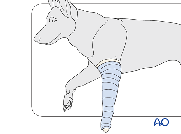
Following appropriate internal fixation, there should be no absolute need for external coaptation for a 23-A2 fracture. If bandaging is used to decrease edema and protect the surgical wound, it should be removed after 24–48 hours to reduce the risk of complications.
Integrative medical therapies, anti-inflammatory medications, and analgesics are recommended.
10–20 minutes of ice therapy is recommended every 8 hours in most cases.
The patient can immediately bear weight, but with strict control of activities.
Phase 2: 4–10 days after surgery
The aim is to resolve hematoma and edema, control pain, and prevent muscle contracture. Anti-inflammatory and analgesic medications may still be needed.
Rehabilitation and integrative medical therapies can be used.
A careful evaluation is recommended if the patient is not starting to use the limb within a few days after surgery.
Phase 3: > 10 days after surgery
The sutures are removed 10–14 days after surgery.
Immature patients should be radiographed every 4 weeks.
A radiographic assessment is performed every 4–6 weeks until bone healing is confirmed, more frequent assessment may be necessary for skeletally immature patients.


