Intralesional resection (S1 to S5)
1. Introduction
Preoperative management
Proper planning is instrumental in the management of primary spine tumors. A multidisciplinary approach may be required depending on the localization of the tumor.
This picture shows a left S1 tumor.

Embolization
Embolization procedures are recommended to reduce operative blood loss in hypervascular tumors, especially during more extensive resections.
Embolization should be considered for hypervascular tumors, such as giant cell tumors, aneurysmal bone cysts, and hemangiomas.
The role of the embolization is:
- To reduce the vascularity of the tumor
- To facilitate dissection around the tumor
- Mapping of spinal cord vascular supply
Embolization on its own may also have a therapeutic effect.
This image shows the embolization of a hypervascular tumor.

Resection strategy
Most benign primary tumors will be localized in the posterior element with variable extension anteriorly. These tumors are approached and resected through a posterior approach only.
A wide visualization is essential in these cases. An adequate amount of sacral lamina needs to be removed to achieve:
- Good visualization of normal and abnormal anatomy
- Safe decompression of the neural elements.
An intralesional approach is preferred for benign tumors, and when an en bloc resection would result in unacceptable morbidity.

Reconstruction strategy
The need for reconstruction will be determined by the amount of bony destruction caused by the tumor.
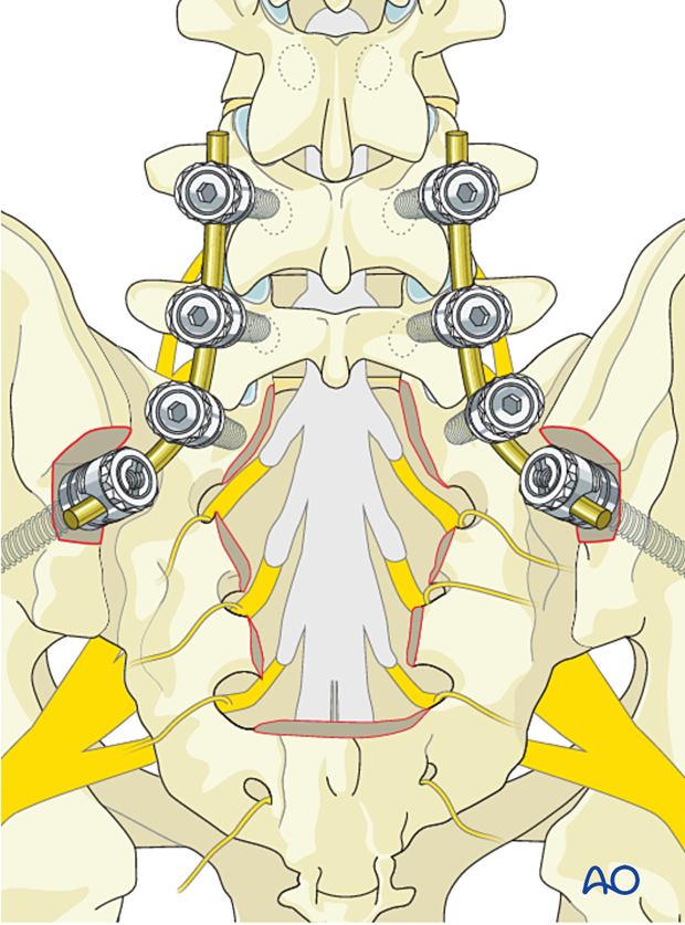
Reconstruction is typically required when more than 50% of the sacroiliac joints are compromised. When reconstruction is required, lumbopelvic fixation is preferred. See En bloc resection (S1-S2).
A classic lumbopelvic reconstruction on the setting of a tumor will be L3 or L4 to the pelvis.
The risk of implant failure may be decreased by cement augmentation of fenestrated screws.
As the procedure is often curative, it is important to verify that the spine is reconstructed in good alignment, and a solid bony union should be attempted.
2. Patient preparation and surgical access
Patient preparation
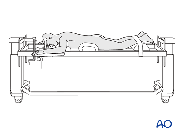
The patient is placed prone on a Jackson table.
Surgical access
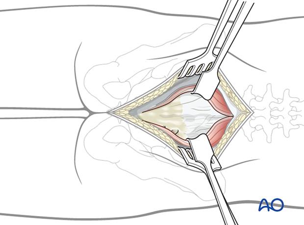
A posterior midline approach to the sacrum is performed.
A wider dissection will typically be performed for primary tumors compared to a trauma case.
3. Sacral laminectomy
Remove intact sacral lamina according to the preoperative planning using a high-speed burr or a Kerrison rongeur.

4. Tumor resection
Dissection of the tumor should progress from normal to abnormal tissues to protect normal neurological elements and facilitate dissection.
Use reverse-angle curettes and pituitary rongeurs to debulk the tumor.
Nerve root mobilization should be minimized to reduce the risk of neurological injury.
The goal is to achieve gross total resection.
Intraoperative navigation can be used as an adjunct to maximize resection accuracy.
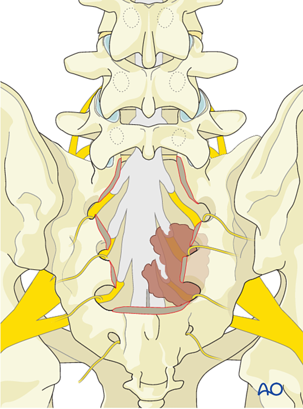
Once the tumor is entirely resected, the goal of the surgery is met.

5. Intraoperative imaging
Before wound closure, intraoperative imaging is performed to check the adequacy of reduction, position, and length of screws, and the overall coronal and sagittal spinal alignment.
6. Posterior closure
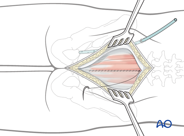
Perform a multilayer closure as described in the approach.
Even a low sacrectomy has a high chance of deep wound infection.
To decrease the risk of wound infections, a gluteal advancement flap may be considered. This is typically performed by the plastic surgeon.
Intrawound vancomycin can be applied to decrease the risk of postoperative wound complications.
7. Aftercare
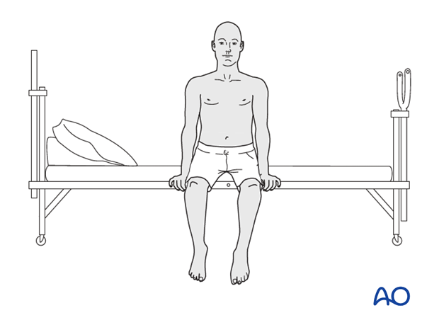
Patients are made to sit up in the bed on the first day after surgery, unless contraindicated because of a plastic flap closure. Patients with intact neurological status are made to stand and walk on the first day after surgery.
Patients can be discharged when medically stable or sent to a rehabilitation center if further care is necessary.
Throughout the hospital stay, adequate caloric intake of a high-quality diet should be monitored.
Patients are generally followed with periodical x-rays at 6 weeks, 3 months, 6 months, and 1 year to monitor for hardware failure and with an MRI every 6 months for tumor surveillance.
Some primary benign tumors of the spine can recur years after surgery, and long-term tumor surveillance is important.


