En bloc resection (S1 to S2)
1. Introduction
En bloc resection of a primary tumor is a significant undertaking, even in the most experienced hands. We therefore recommend referring these cases to quaternary centers with experience in primary spine tumor surgery.
En bloc resections
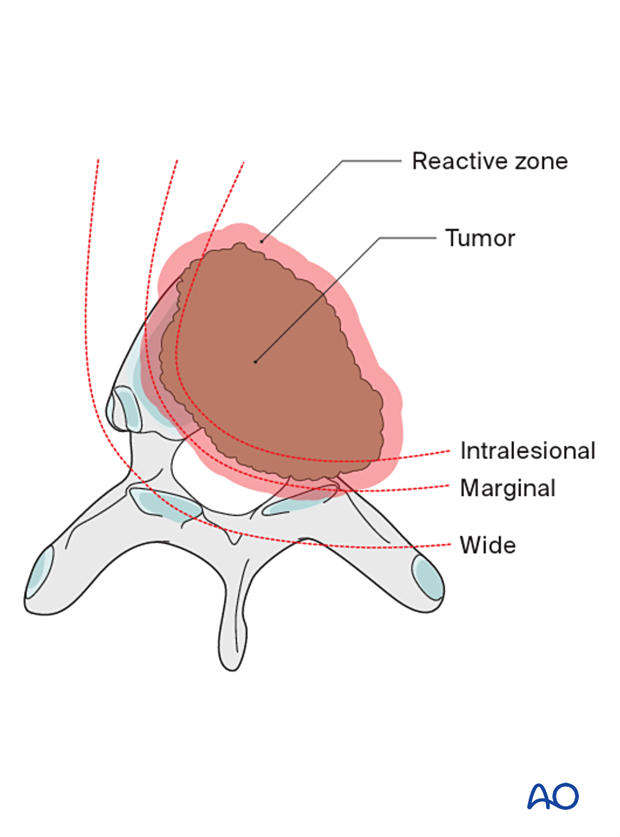
Terminology is essential in primary tumor management.
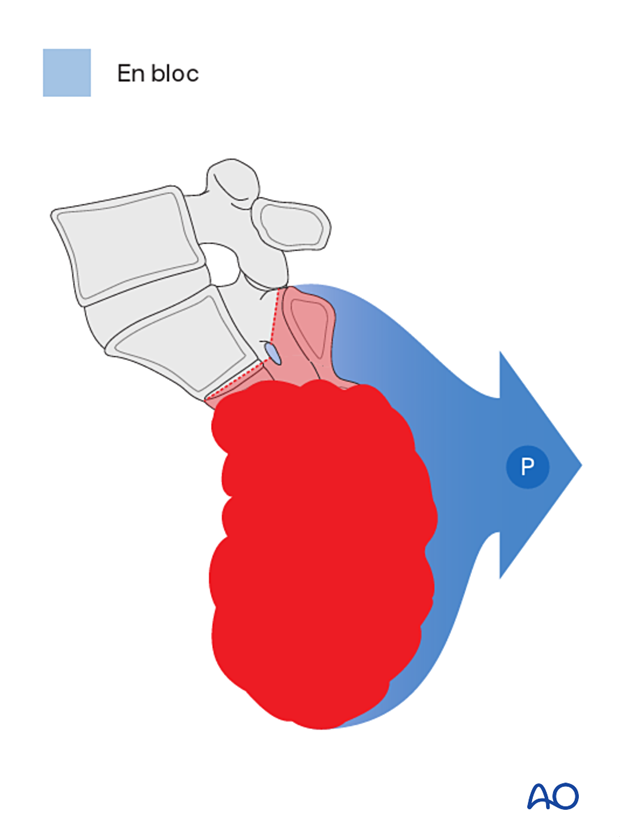
An en bloc resection refers to a surgical attempt to remove a tumor in one piece without violating it.
On the other hand, an intralesional resection or a curettage refers to a deliberate intralesional resection.
An en bloc resection needs to be associated with a pathological margin description to be correctly defined.
Four types of margins are described:
- Intralesional – resection margin is within tumoral tissue
- Marginal – resection margin is within a reactional zone or pseudocapsule (in the spine, the epidural margin is often marginal)
- Wide – resection margin is within normal tissue
- Radical – this is extracompartmental resection and, as such, does not apply to spine tumors
2. Planning
Preoperative management
Proper planning is instrumental in the management of primary spine tumors. A multidisciplinary approach may be required depending on the localization of the tumor.
A CT scan, an MRI, and a spinal angiogram will assist in the final planning. 3D models, if available, can also help in the planning of those cases.

Embolization
Embolization procedures are recommended to reduce operative blood loss in hypervascular tumors, especially during more extensive resections.
Embolization should be considered for hypervascular tumors, such as giant cell tumors, aneurysmal bone cysts, and hemangiomas.
The role of the embolization is:
- To reduce the vascularity of the tumor
- To facilitate dissection around the tumor
- Mapping of spinal cord vascular supply
Embolization on its own may also have a therapeutic effect.
This image shows the embolization of a hypervascular tumor.

Preoperative planning
For high sacrectomies, both an anterior and posterior approach are required.
During the initial anterior approach, the goals are:
- Anterior tumor dissection from the rectum and vascular structures
- Consideration of a diversion colostomy
- Vertical Rectus Abdominus Muscle (VRAM) flap harvest
During the posterior approach, the osteotomies are completed, and the tumor is delivered.
A wide visualization is essential in these cases. An adequate amount of sacral lamina needs to be removed to achieve:
- Good visualization of normal and abnormal anatomy
- Safe decompression of the neural elements.
Sacral nerve root sacrifice is associated with neurological morbidity.
If at least one S3 nerve root is preserved, a degree of continence will be maintained in most patients.
For a high sacral resection (as in this case), the bilateral S2 to S5 nerve roots will be sacrificed. Complete loss of bowel, bladder and sexual function can be expected.
A diversion colostomy may be considered to reduce the risk of wound complications.
A multidisciplinary team is needed to perform a sacrectomy. This team should include a spine surgeon, a plastic surgeon, and a general surgeon. A vascular surgeon may also be needed.
This picture shows a large sacrum chordoma.
Resection strategy
If reconstruction is required, then a lumbopelvic reconstruction is performed.
The general indication to perform a lumbopelvic reconstruction is the removal of more than 50% of the SI joint.
Case-based scenario
Every case is unique.
To illustrate the surgical principles of a staged anterior/posterior sacrectomy, we will use a high sacral tumor located in S2–S5. To resect this tumor, more than 50% of the sacroiliac joint will be resected, and a lumbopelvic reconstruction will be performed.

3. Anterior approach
Patient preparation
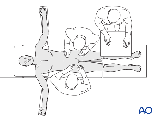
The patient is placed supine on a Jackson table.
Surgical approach
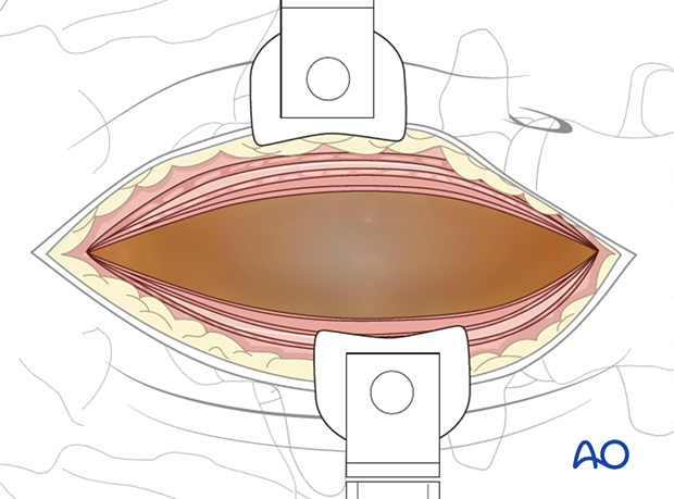
An access surgeon is required to perform the anterior retroperitoneal approach.
In principle, this approach is an extended version of the mini open approach.
Anterior release of the tumor
The access surgeon mobilizes the rectum and vascular structures away from the tumor.
The plastic surgery team harvests a VRAM flap used during the posterior approach.
If a diversion colostomy is planned, this is now performed.
The wound is closed in a multilayer fashion.

4. Posterior approach
Patient position
The patient is turned and placed prone on a Jackson table. Regular prone positioning or a split leg attachment can be used. The potential advantage of a split leg attachment is that a third surgeon can work between the legs to connect the posterior and anterior exposure.

Surgical access
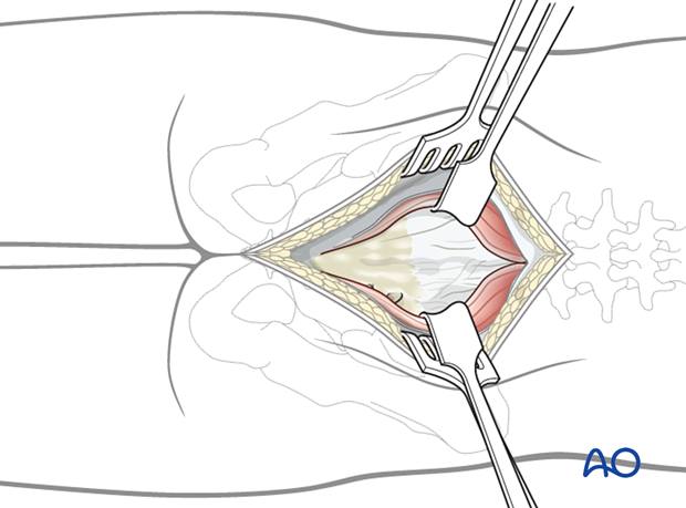
A posterior midline approach to the sacrum is performed.
A wider dissection will typically be performed for primary tumors compared to a trauma case.
Great care should be taken not to enter the tumor during exposure.
Review preoperative images to verify whether the tumor invades the lamina. In such cases, exposure of the posterior elements should be performed with great care, and the use of Cobb elevators should be avoided.
5. Lumbosacral stabilization
Pedicle screw insertion
In the case of a complete sacrectomy or when more than 50% of the SI joints are removed, lumbopelvic reconstruction is performed and will be described here.
Insert all screws according to the preoperative plan.
Optimal pedicle screw purchase will, in order of importance, be achieved by:
- Selecting the largest possible screw diameter
- Selecting the longest possible screw
- Positioning of the screw under the cranial endplate
- Cement augmentation of the screw.

Lumbar and S1 pedicle screws are inserted according to the standard technique.

Iliac screw insertion
Iliac screws are inserted in one of the three potential locations.
For complex lumbopelvic reconstruction, dual iliac screws should be considered.
The iliac screw heads and connecting rods must lie flush with the bone. Avoiding prominent hardware prevents soft tissue irritation.
With a sacral entry point for the iliac screw, there is less concern with screw prominence compared to an iliac starting point.

Rod preparation
Cut the rods to the appropriate length. Every effort should be made to contour the rod to decrease the risk of induced sagittal or coronal malalignment.

6. Posterior delivery
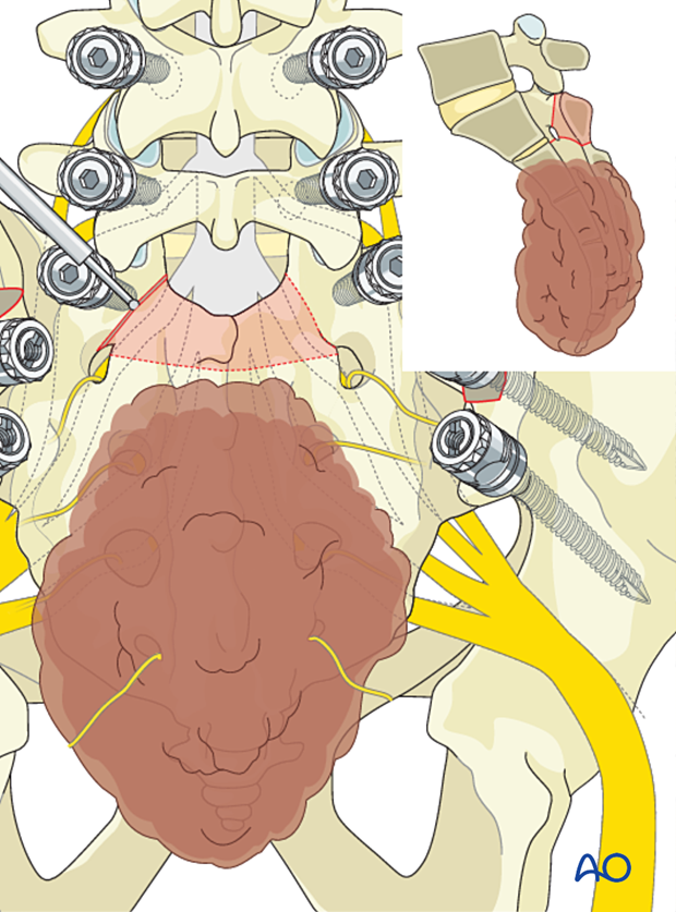
Sacral laminectomy
Remove intact sacral lamina according to the preoperative plan using a high-speed burr or a Kerrison rongeur.
Dissection should progress from normal to abnormal tissues to protect normal neurological elements and facilitate dissection.
Soft-tissue dissection
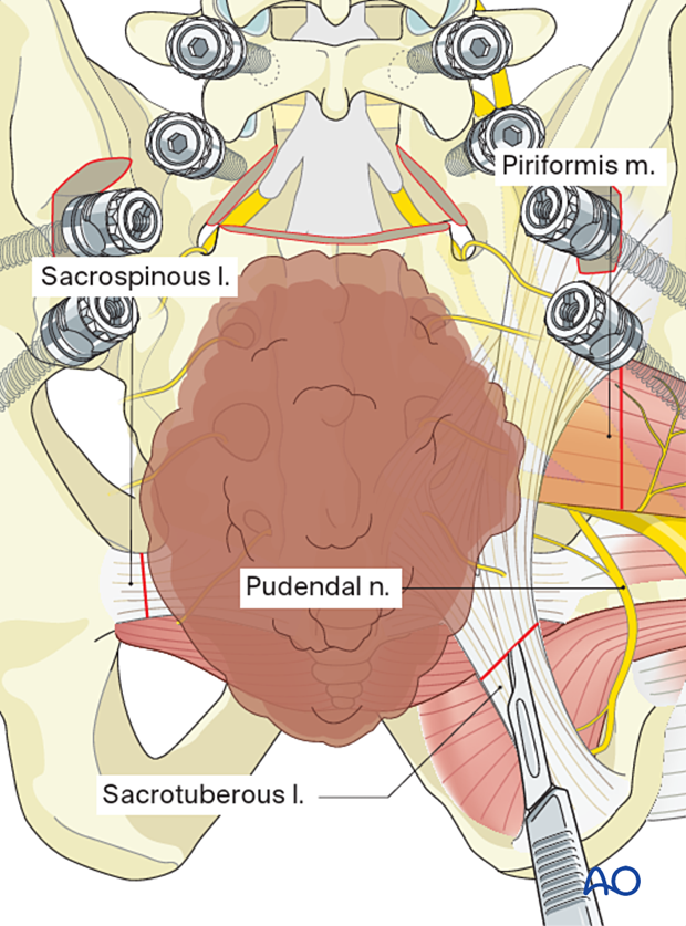
Dissect the posterolateral soft tissues around the tumor in normal tissue planes.
The goal is to join the ventral dissection planes created during the anterior approach.
Divide the sacral tuberous ligaments and the sacral spinous ligaments.
To decrease the risk of local recurrence, ensure a liberal piriformis muscle margin.
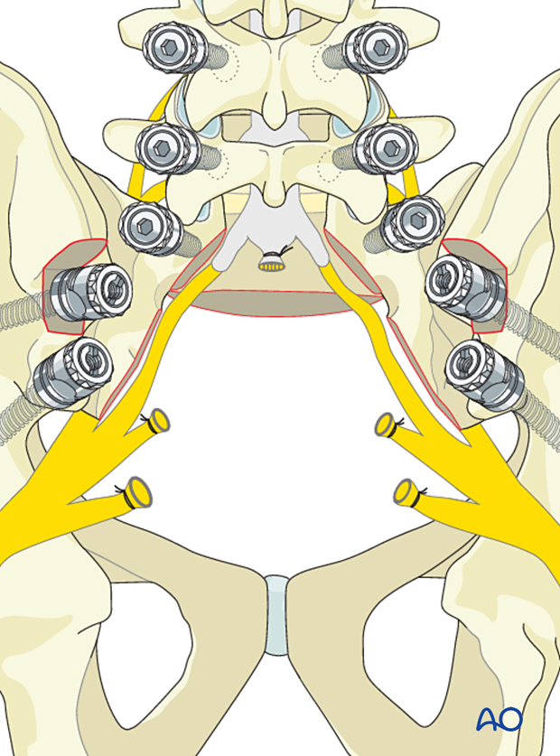
Thecal sac ligation
Ligate the thecal sac at the chosen level.
In this case, just distal to the S1 nerve root takeoff.

Remove the bone (shadowed in the picture) by following the S1 nerve root out laterally to visualize and protect it.
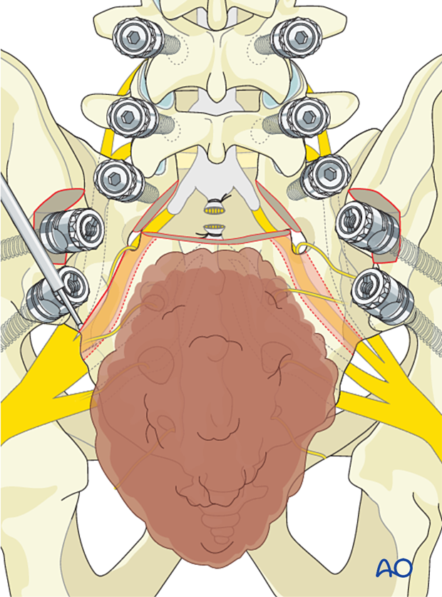
With an osteotome or bone scalpel, perform the transverse osteotomy of the anterior sacrum, between the S1 foraminae, just distal to the nerve root, as illustrated.

Tumor delivery
Deliver the tumor.
7. Fusion
Preparation for fusion
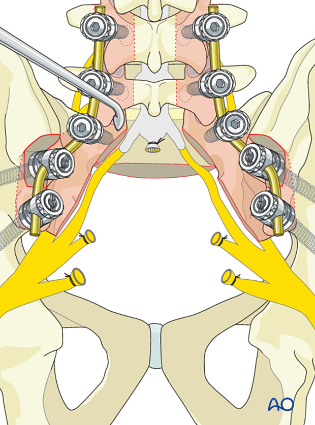
Attach the rods to the screws with the hardware system. Then excise the facet capsule and denude/curette the joint surface cartilage surfaces and the posterior cortex.
Grafting
Insert pieces of bone graft (autograft, allograft) into the decorticated facet joint for fusion.

8. Intraoperative imaging
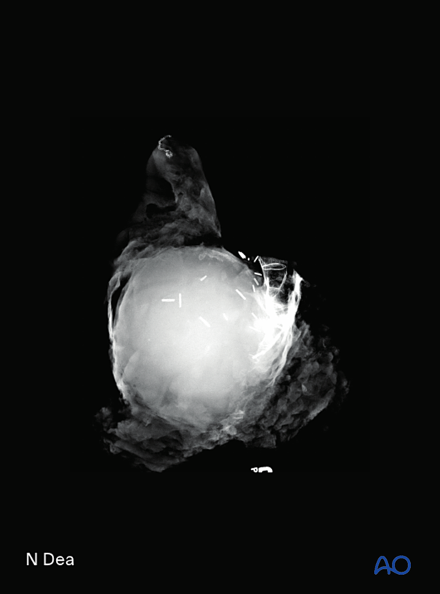
Lateral view of a specimen of an S2–S5 sacral chordoma resection.
9. Posterior closure
The VRAM flap is retrieved from the pelvis and fixed in place to fill the soft tissue defect.
A mesh could be considered to decrease the risk of a dorsal hernia.
Multilayer closure is performed by, or with the assistance of, the plastic surgery team.
10. Aftercare
Patients are mobilized progressively without any direct pressure on the wound as directed by the plastic surgeon.
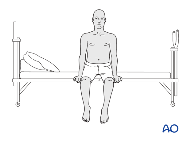
Patients with intact neurological status are made to stand and walk on the first day after surgery.
After a high sacrectomy, bowel and bladder deficits are to be expected, and appropriate rehabilitation should be provided.
Patients can be discharged when medically stable or sent to a rehabilitation center if further care is necessary.
Throughout the hospital stay, adequate caloric intake of a high-quality diet should be monitored.
Patients are generally followed with periodical x-rays at 6 weeks, 3 months, 6 months, and 1 year to monitor for hardware failure and with an MRI every 6 months for tumor surveillance.
Some primary benign tumors of the spine can recur years after surgery, and long-term tumor surveillance is important.


