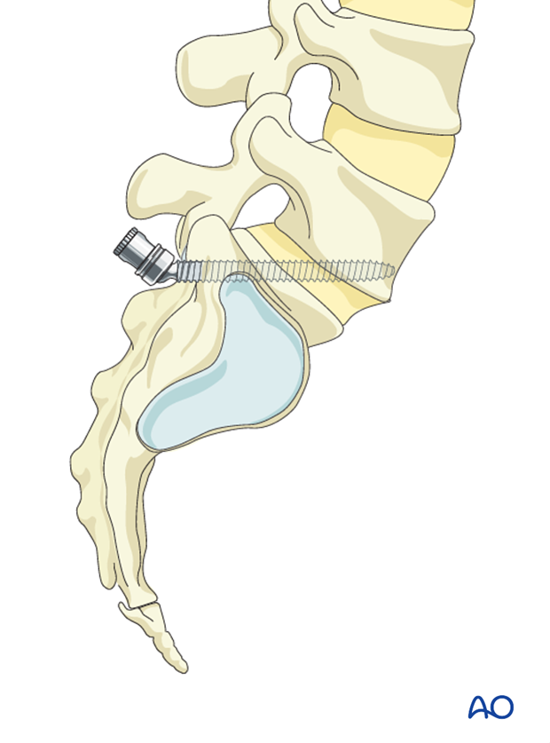
S1 Pedicle and delta screw insertion
1. Introduction
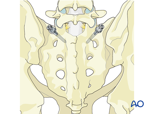
Pedicle screws in S1 may be used in the management of trauma, deformities, tumors, and degenerative conditions of the lumbar and sacral spine.
For an alternative fixation technique (delta fixation), a more cranial oriented trajectory can be used.
2. Preparation
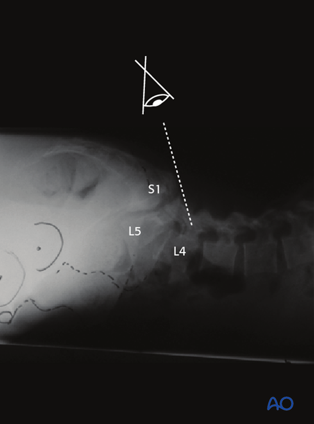
Once the spine is exposed, the appropriate levels of fixation are confirmed with the image intensifier.
3. Pitfall in spondylolisthesis
Due to the distorted anatomy care must be taken to confirm correct fusion levels. Typically, the L5 pedicle is extremely anterior, hidden beneath the sacral alar.
In high grades it recommended to span the fusion from L4 to S1 or pelvis.
4. Entry points
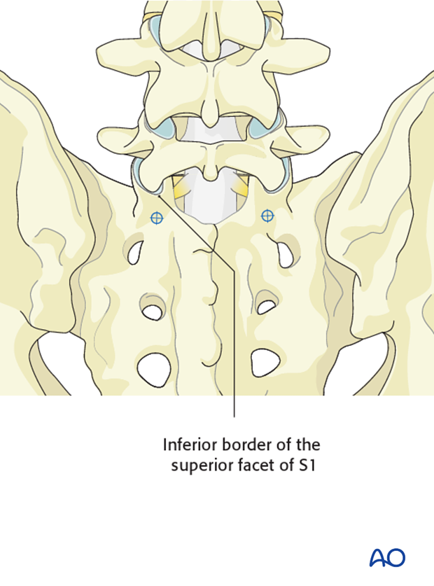
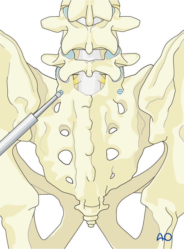
The entry point of the pedicle screw is defined by the inferior border of the superior facet of S1.
5. Opening of the cortex
Open the superficial cortex of the entry point with a burr or a rongeur.
6. Medio-lateral inclination
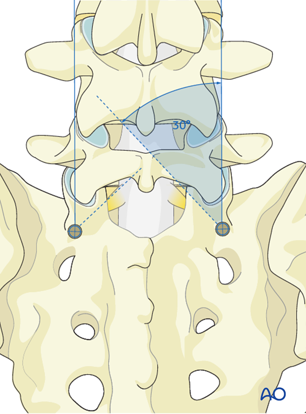
The trajectory of the screw is 30° medially.
7. Cranial caudal angulation
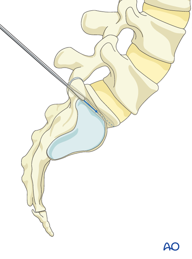
A pedicle probe is used to navigate down the isthmus of the pedicle into the vertebral body. The appropriate trajectory of the pedicle probe in the cranial caudal direction occurs by aiming towards the promontory.
Lateral X-rays are taken to confirm tip of probe end in promontory of S1.
8. Screw insertion
Once the pedicle track has been created, it is important to confirm a complete intraosseous trajectory by pedicle and body palpation using a pedicle sounding device. At any point in the process, radiographic confirmation can be obtained.
Note: The selection of a mono- or a polyaxial screw is usually the choice of the surgeon.
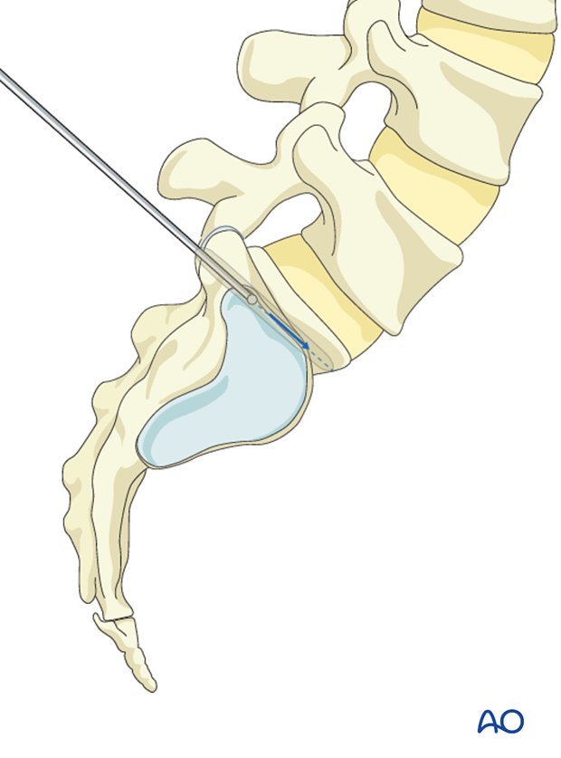
A screw of appropriate diameter and length is carefully inserted into the same created trajectory.
Classically these screws are large in diameter, however, shorter in length than the L5 screws. If screws are too long and breach the anterior cortex, L5 nerve root and great vessels are at risk of injury.
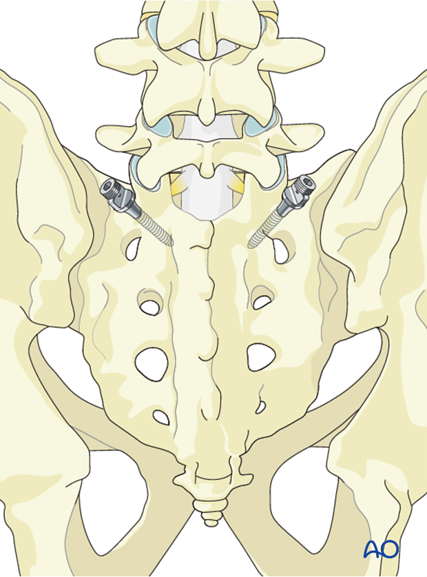
9. Screw insertion for delta fixation
For an alternative fixation technique (delta fixation), a more cranial oriented trajectory can be used.
This technique can be used in selected cases of Type 5 where reduction is not necessary, but added stability is desired.
The entry point remains the same as the standard S1 pedicle screw.
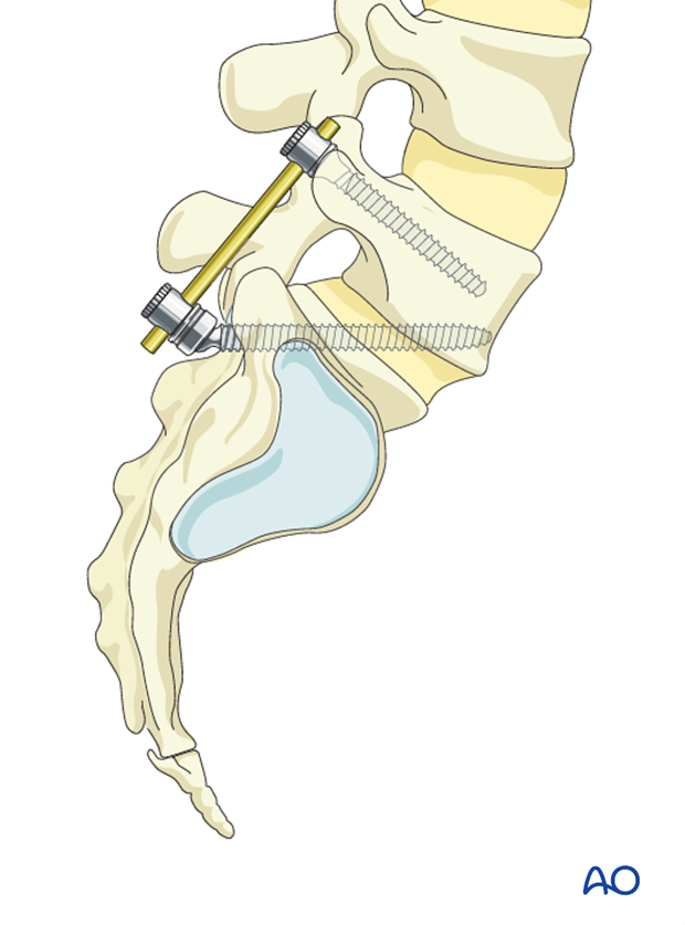
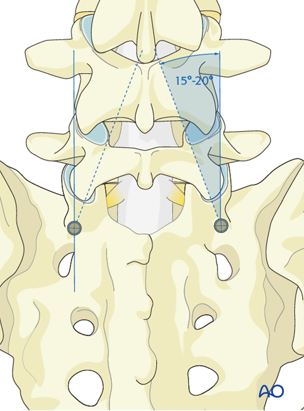
The medial inclination is roughly 15°–20°.
The cranial angulation must allow for perforation of the superior endplate of S1 crossing the L5–S1 and the inferior endplate of L5 into the body of L5.
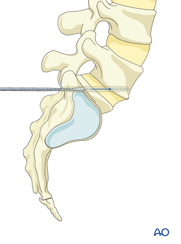
This trajectory requires a longer screw than the standard S1 screw. Screw diameter should be 6mm or more.
