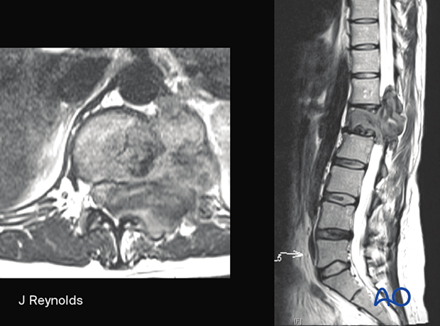
New module - Spine primary tumors
Introduction
Publication of first edition (May 2023)
The first edition was created by Nicolas Dea (Canada), and Jeremy Reynolds (UK), with Luiz Vialle (Brazil) as editor.
This module is based on current clinical principles, practices, and available evidence, and supports spine surgeons’ day-to-day treatment, planning, learning, and teaching. The module joins existing content on metastatic lesions and completes the AO Surgery reference chapter on tumors.
Visit the module entry page here!

Highlights
The new module of primary tumors details treatments tailored to benign and malignant tumors in various segments of the spine. Additionally, the module offers tumor definitions, treatment indications, guides to patient positioning, and approaches.
The module addresses:
A wide range of basic techniques are also described.
Content
Table of contents
The Primary tumors module includes a detailed description of the following tumor types and their management.
Benign tumors (Osteoid osteoma, Osteoblastoma, Giant cell tumors, Aneurysmal bone cyst, Hemangioma)
C1–C7:
- Observation and analgesics
- Chemotherapy
- Radiofrequency ablation
- Percutaneous treatment (embolization)
- Radiation therapy
- Intralesional resection (C0–C2)
- Intralesional resection (C3–C7)
- En bloc resection of an anterior tumor (C1–C7)
- En bloc resection of a posterior tumor (C1–C7)
T1–T12
- Observation and analgesics
- Chemotherapy
- Radiofrequency ablation
- Percutaneous treatment (embolization)
- Radiation therapy
- Intralesional resection (L1–L5)
- En bloc resection with posterior release and anterior tumor delivery (L1–L5)
- En bloc resection of a posterior tumor (L1–L5)
L1–L5:
- Observation and analgesics
- Chemotherapy
- Radiofrequency ablation
- Percutaneous treatment (embolization)
- Radiation therapy
- Intralesional resection (L1–L5)
- En bloc resection with posterior release and anterior tumor delivery (L1–L5)
- En bloc resection of a posterior tumor (L1–L5)
S1–S5:
- Observation and analgesics
- Chemotherapy
- Radiofrequency ablation
- Percutaneous treatment (embolization)
- Radiation therapy
- Intralesional resection (S1–S5)
- En bloc resection (S1–S2)
- En bloc resection (S3–S5)
Malignant tumors (Chondrosarcoma, Osteosarcoma, Ewing's sarcoma, Chordoma)
C1–C7:
- Radiation therapy
- Neoadjuvant therapy
- En bloc resection of an anterior tumor (C1–C7)
- En bloc resection of a posterior tumor (C1–C7)
- Adjuvant therapy
- Palliative care
T1–T12:
- Radiation therapy
- Neoadjuvant therapy
- En bloc resection of an anterior tumor (T1–T12)
- En bloc resection of a posterior tumor (T1–T12)
- Adjuvant therapy
- Palliative care
L1–L5
- Radiation therapy
- Neoadjuvant therapy
- En bloc resection with posterior release and anterior tumor delivery (L1–L5)
- En bloc resection of a posterior tumor (L1–L5)
- Adjuvant therapy
- Palliative care
S1–S5
- Radiation therapy
- Neoadjuvant therapy
- En bloc resection (S1–S2)
- En bloc resection (S3–S5)
- Adjuvant therapy
- Palliative care
Additional material
Approaches:
- Posterior access to the occipitocervical spine (C0–C2)
- Posterior access to the cervical spine (C3–C7)
- Anterior access to the cervical spine (C3–C7)
- Posterior access to the cervicothoracic junction (C7–T2)
- Posterior midline access to the thoracolumbar spine (T1–L5)
- Retroperitoneal approach (L4–S1)
- Posterior midline approach to the sacrum (S1–S5)
Patient preparations:
- Prone position for approaches to C0–C7
- Supine position for approaches to C3–C7
- Prone position for approaches to T1–S5
- Supine position for approaches to T1–S5
Further reading:
- Essential initial patient assessment
- Neurological examination
- Oncological and surgical staging
- Sagittal spinal alignment
- References on primary spine tumors
Basic techniques:
- Ligation of nerves
- Instrumentation for occipitocervical fixation
- Cervical discectomy
- PMMA application for vertebral body reconstruction
- Pedicle screw insertion (T1–T3)
- Pedicle screw insertion in the cervical spine
- Lateral mass screw insertion (Magerl technique)
- C1 lateral mass screw insertion
- C2 pedicle screw insertion
- C2 pars screw insertion
- C2 laminar screw insertion
- Pedicle screw insertion in the thoracolumbar spine
- S1 pedicle screw insertion
- Iliac screw insertion


