Posterior long segment fixation
1. Introduction
Preliminary remarks
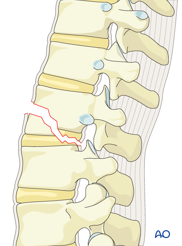
B3 injuries are commonly seen in ankylotic disorders such as diffuse skeletal hyperostoses and ankylosing spondylitis. It is a hyperextension injury through the disk or vertebral body leading to a hyperextended position of the spinal column. Anterior structures, notably the anterior long ligaments (ALL) are ruptured.
Repair of dural laceration
More details on repair of dural laceration can be found here.
2. Patient preparation and approach
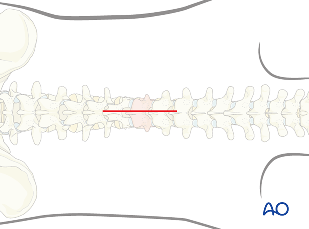
The posterior open approach to the midline is used together with the appropriate patient preparation.
3. Closed reduction
Primary reduction is performed by positioning of the patient onto a frame to create lordosis.
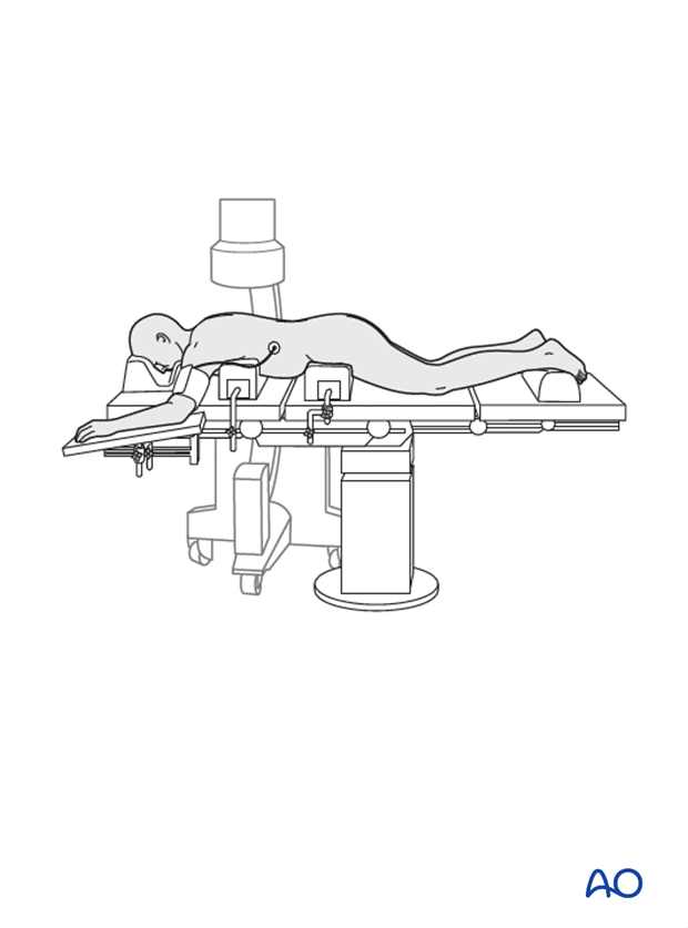
4. Reduction with pedicle screws
Preliminary remarks
Due to the fact that bilateral instrumentation is necessary in all cases, all steps described below are repeated on the opposite side, unless stated otherwise.
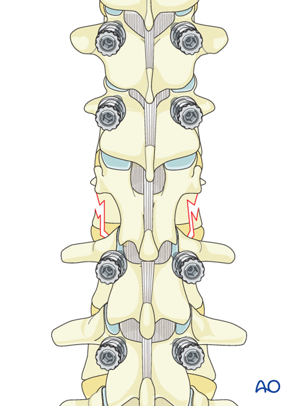
Pedicle screw insertion
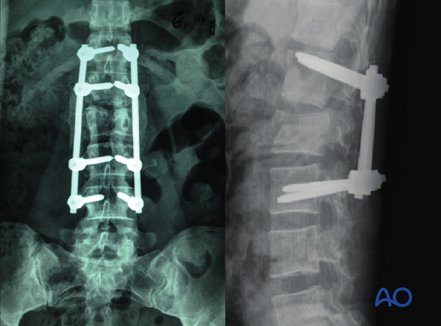
Since B3 injuries occur in a stiff spine, posterior long segment fixation is performed to provide better reduction forces and biomechanical stability. Short segment constructs lead to increased stress on the posterior implants resulting in implant failure. B3 fractures associated with significant comminution of the vertebral body (A3, A4) also may require anterior reconstruction to achieve better biomechanical stability. If the vertebral body fracture is of A1 or A2 types, then posterior long segment fixation alone suffices.
In this technique, pedicle screws are inserted two levels above and below the fractured level on both sides.
This is essential in rotationally unstable injuries where stress on the posterior implants is high.
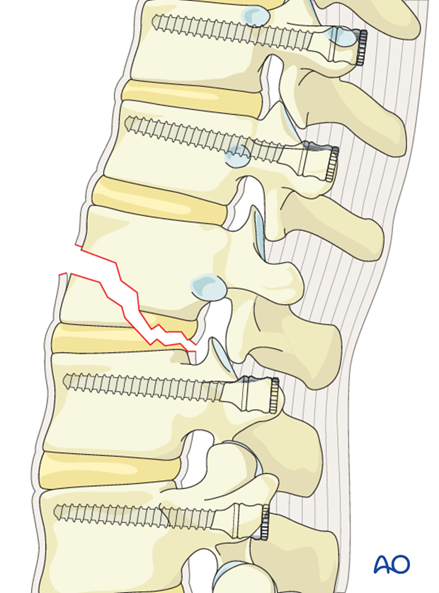
Rod contouring
The contouring of the rod depends on the site of the fracture. A rod contoured in mild kyphosis is chosen for fractures from T1-T10. A straight or a slightly lordotic rod is chosen for fractures from T11-L1, and a rod contoured to lordosis is chosen for lumbar fractures.
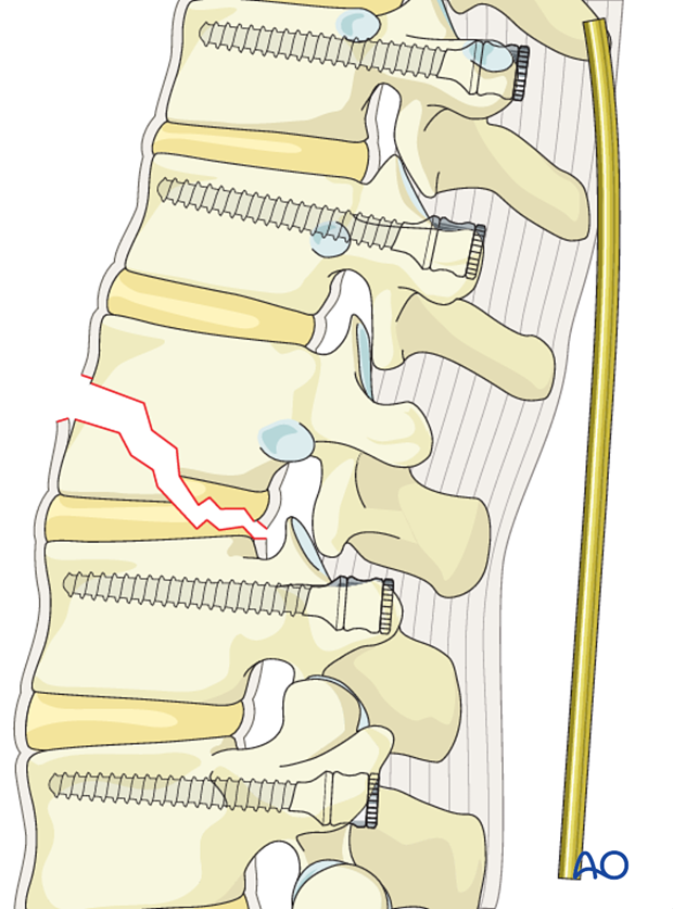
Rod insertion
The rod is inserted sequentially, either from distal to proximal or the other way round. The distal screw heads are tightened.
The rod is then inserted into the proximal screw heads without tightening.
Proper rod contouring helps in achieving appropriate sagittal alignment.
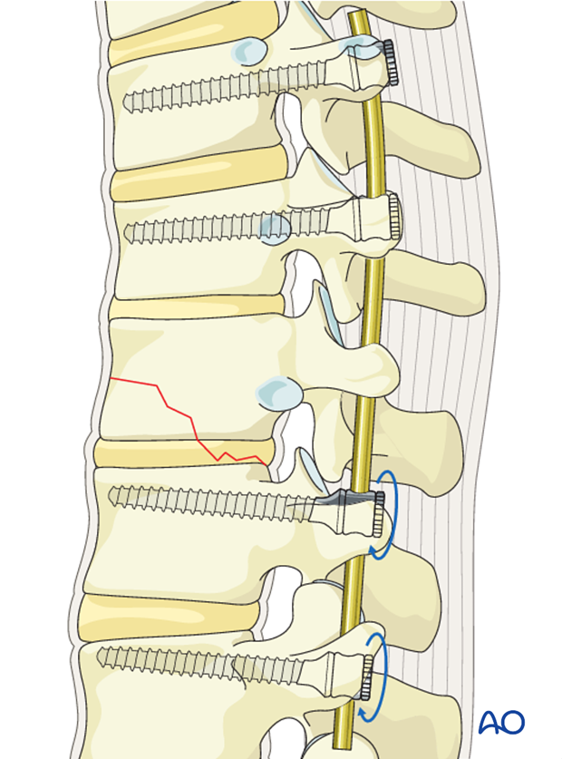
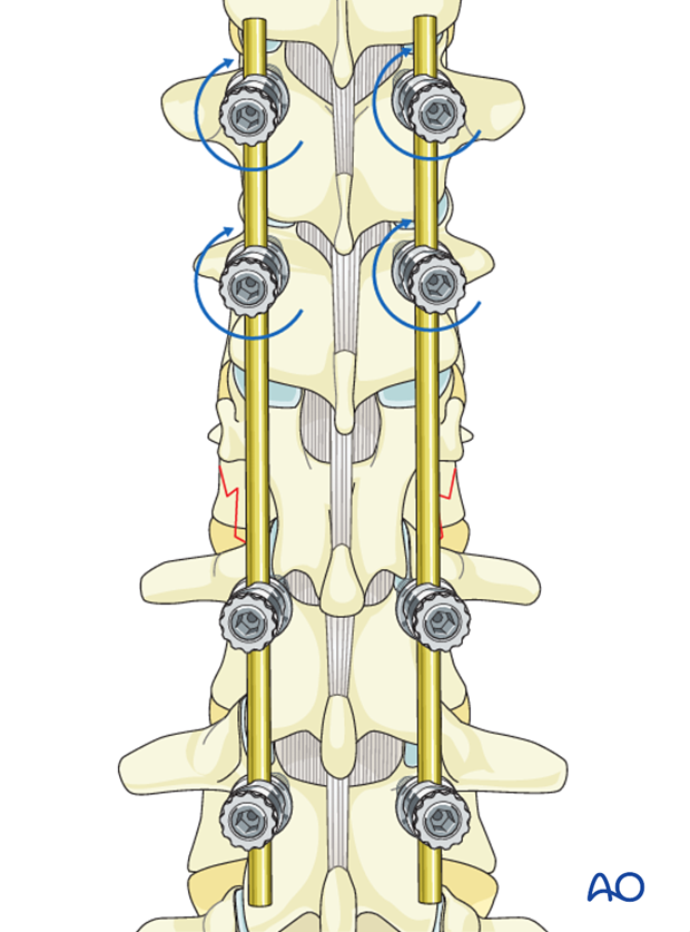
The screw heads are tightened with the inner nuts to secure the reduction achieved.
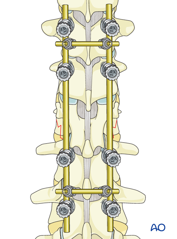
Cross links are applied at the top and bottom of the construct to provide biomechanical stability. In type A injuries one cross link might be sufficient.
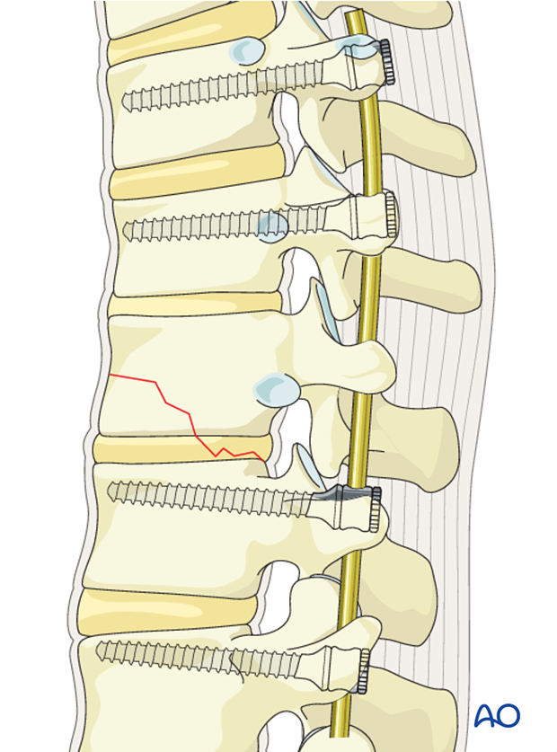
The final construct is shown from a lateral view
5. Fusion
Decision
Although fusion was routinely performed for all spinal fractures, its indications are now being restricted to fractures that are highly unstable.
Nonfusion fixations can be performed for A3, A4, and B1 type injuries.
Fusion is routinely performed for A2, B2, B3, and all C injuries as they are unstable injuries with extensive soft tissue and ligamentous disruption.
If the surgeon plans for a fusion, the facet capsule is excised and the joint cartilage surfaces are denuded/curetted.
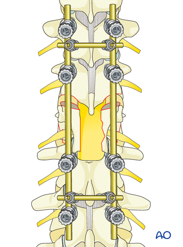
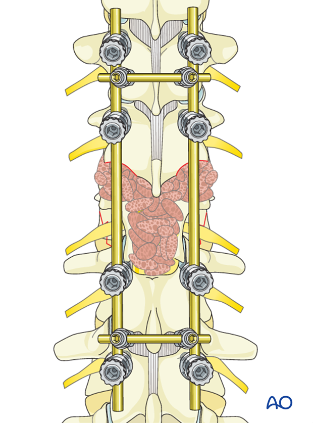
Pieces of bone graft (autograft, allograft) are inserted into the decorticated facet joint for fusion.
6. Intraoperative imaging
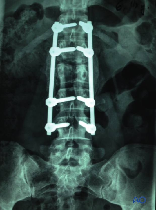
Prior to wound closure, intraoperative imaging is performed to check the adequacy of reduction, position, and length of screws and the overall coronal and sagittal spinal alignment.
7. Aftercare for posterior procedures
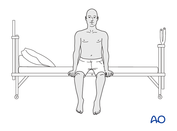
Patients are made to sit up in the bed on the first day after surgery. Bracing is optional. Patients with intact neurological status are made to stand and walk on the second day after surgery. Patients can be discharged when medically stable or sent to a rehabilitation center if further care is necessary. This depends on the comfort levels and presence of other associated injuries.
Patients are generally followed with periodical x-rays at 6 weeks, 3 months, 6 months, and 1 year. Normally, 5-10 degrees of loss of kyphosis can be observed within the first 6 months, which does not affect the functional outcomes. For nonfusion surgeries, the implants can be removed once fusion is confirmed.
