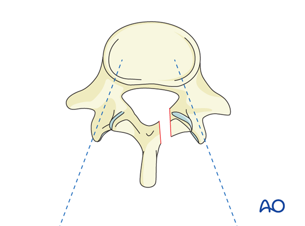
Pedicle screw insertion in the thoracic spine
1. Introduction
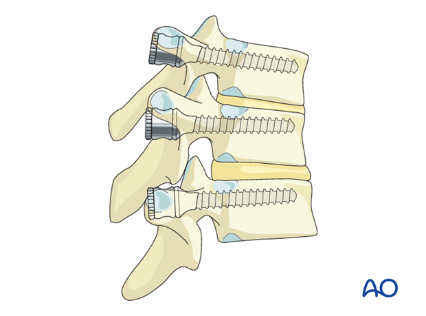
Pedicle screws in the thoracic spine may be used in the management of trauma, deformities, tumors, and degenerative conditions of the thoracic spine and its neighboring segments.
2. Preparation
Once the spine is exposed, the appropriate levels of fixation are confirmed with the image intensifier.
3. Unstable injuries after trauma
In unstable injuries, the segments above and below the level of injury may have a different orientation of pedicle trajectory due to displacement and rotation.
In severely unstable injuries, care must be taken not to cause more displacement while using the pedicle awl.
Whenever necessary, image intensifier confirmation of the starting point and trajectory must be obtained.
Preoperative assessment of pedicle integrity must be done for all levels of instrumentation.
4. Landmarks
Landmarks:
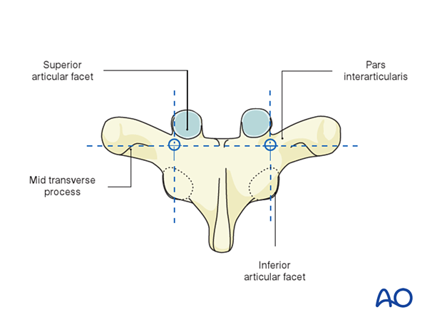
- Lateral border of the superior articular facet
- Lateral border of the inferior articular facet
- Ridge of the pars interarticularis
- Mid transverse process
5. Entry points in the thoracic spine
Pedicle screw entry points and angulation in the thoracic spine can be divided into four groups based on the relation of the pedicle with the articular facets and the transverse process.
The entry point of the pedicle screw for the lower thoracic segments is defined after determining the intersection of the mid portion of the facet joint and the superior edge of the transverse process. The specific entry point will be just lateral and caudal to this intersection.
The entry point tends to be more cephalad as you move to more proximal thoracic levels.
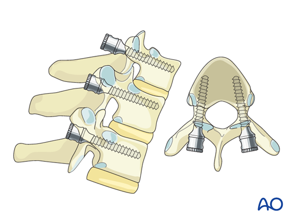
T1–T3
The entry points is in the intersection of the line drawn horizontally though the middle of the transverse process with a line drawn slightly lateral to the center of the articular facet.
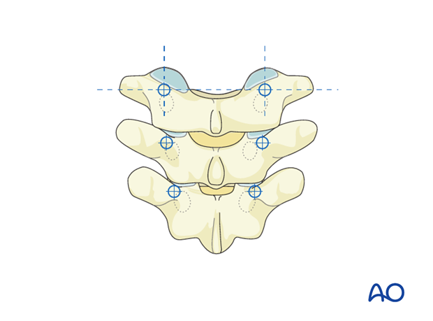
The screw angulation is slightly medial and caudal.
T4–T6
The entry point is more cranial and medial compared to the T1–T3 entry points. The entry point is in the intersection of the line drawn horizontally through the upper 1/3 of the transverse process and the line drawn through the center of the articular facet.
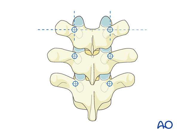
The screw angulation is almost vertical.
T7–T9
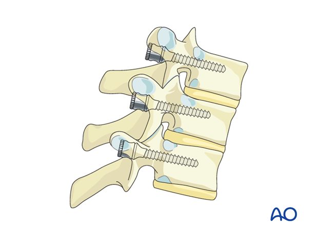
The entry point is more cranial and medial compared to the T4–T6 entry points. The entry point is in the intersection of the line drawn horizontally on the superior border of the transverse process and the line drawn slightly medial to the center line of the articular facet.
The screw angulation is vertical.

T10–T12
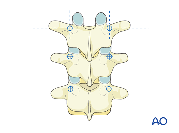
These pedicles tend to be very narrow. The entry is in the intersection of a horizontal line drawn through the mammillary process or the upper 1/3 of the transverse process, and the vertical line drawn through the center of the articular facet.
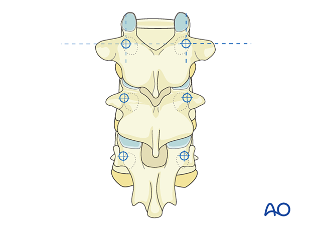
The screw angulation is vertical or slightly lateral.
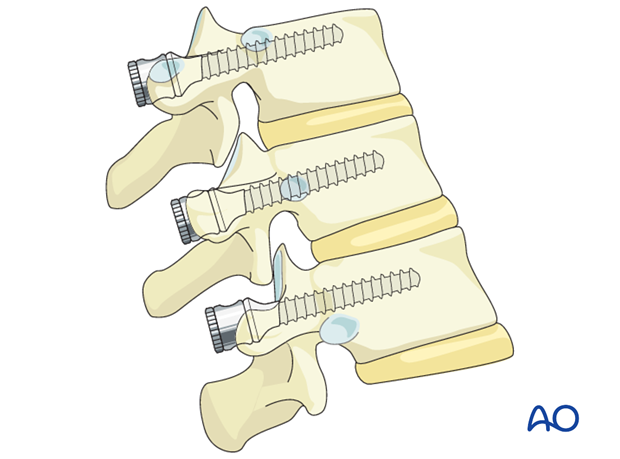
If one is not sure the entry point for the pedicle is correct, a small laminotomy with removal of the yellow ligament will allow palpation of the inner wall of the pedicle with either a pennfield or a ball-tip probe.
