Posterior open approach - midline approach (T1-S1)
1. Skin incision
The skin and subcutaneous tissue is infiltrated with a 1:500,000 epinephrine solution to achieve hemostasis.
A midline skin incision is made centred over the involved segment. The length of the incision depends on the number of levels to be instrumented. For a short segment fixation, one level above and below the fractured vertebra is exposed.
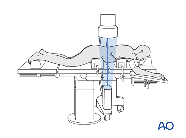
It is necessary to confirm the correct level of the approach with fluoroscopy.
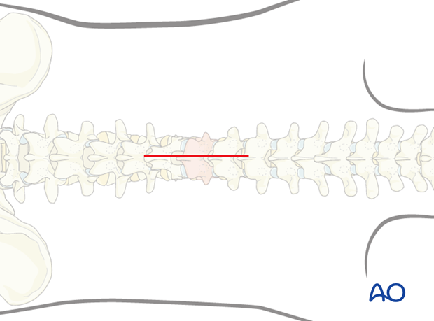
For a long segment fixation, two segments above and below the fractured vertebra is exposed.
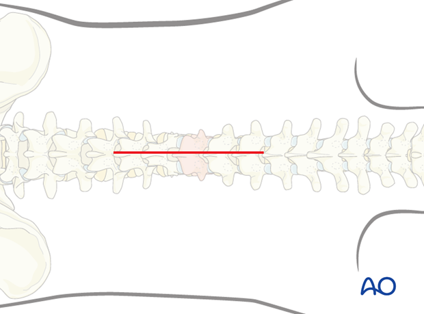
2. Exposure
The dissection is carried down in the midline through the subcutaneous tissue and the fascia to the tips of the spinous processes. Self-retaining retractors are used to maintain tension on soft tissues during exposure.
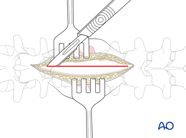
The paraspinal muscles are elevated subperiosteally from the underlying laminae, in distal to proximal direction, using a Cobb elevator. Dissection is done along the spinous process and lamina.
The use of a subperiosteal dissection minimizes bleeding and muscle damage.
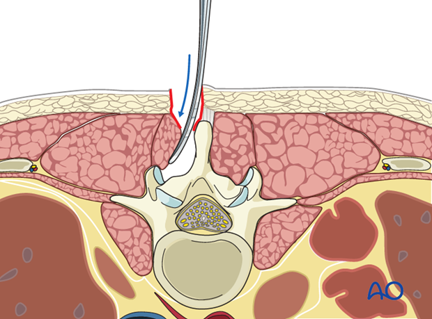
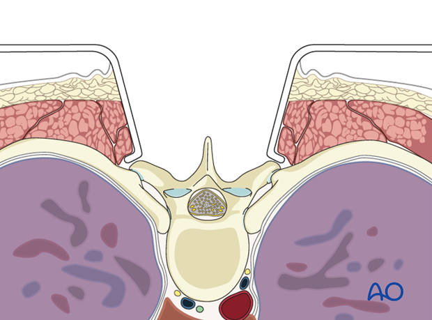
In the thoracic region, the dissection is usually taken to the tips of the transverse processes.
In the lumbar spine, the dissection is limited to the facet joints. The extent of dissection will depend upon the need and type of fusion planned.
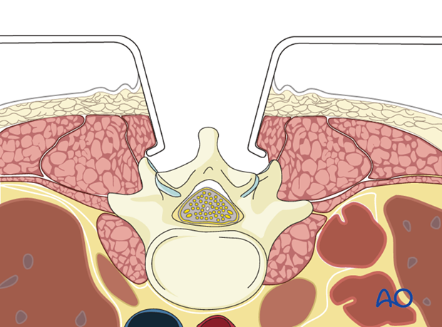
Note: during exposure, care is taken not to injure the facet joint capsule if a nonfusion technique is planned.
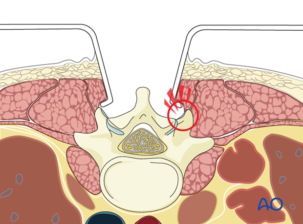
Alternatively, if the surgeon plans for a fusion, the facet capsule is excised and the joint cartilage surfaces are denuded for fusion.
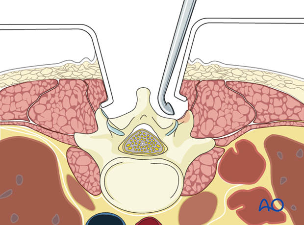
Localizing x-ray or image intensifier check of spinal level should be obtained once exposure is completed.
Pitfall: Laminar split fractures
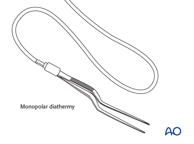
In at least 30% of patients with burst fractures and incomplete neurological deficit, a split fracture of the lamina is present. Dural sheath and or nerve roots can be impaled into this defect. Monopolar diathermy can potentially injure these neural structures if one is not aware. The use of a bipolar cautery is recommended.
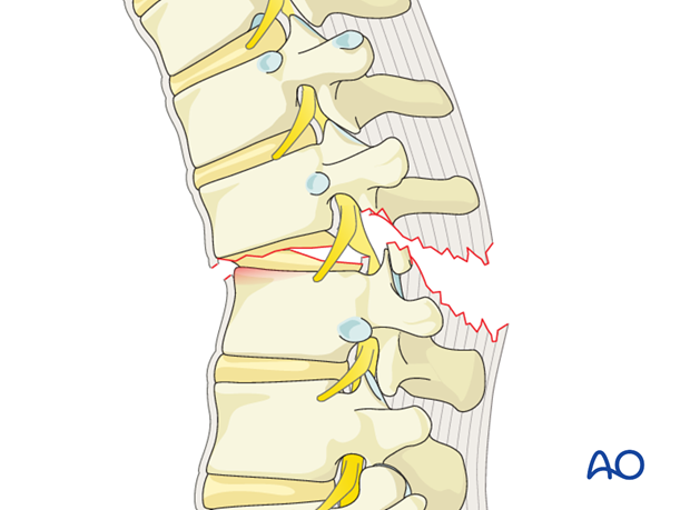
Pitfall: type B1 and B2 injuries
In patients with B1 and B2 injuries, due to posterior distraction injury to the PLC, there can be defects in the ligamentum flavum and lamina. The surgeon should be aware about this potential defect while exposing the spine in such patients.
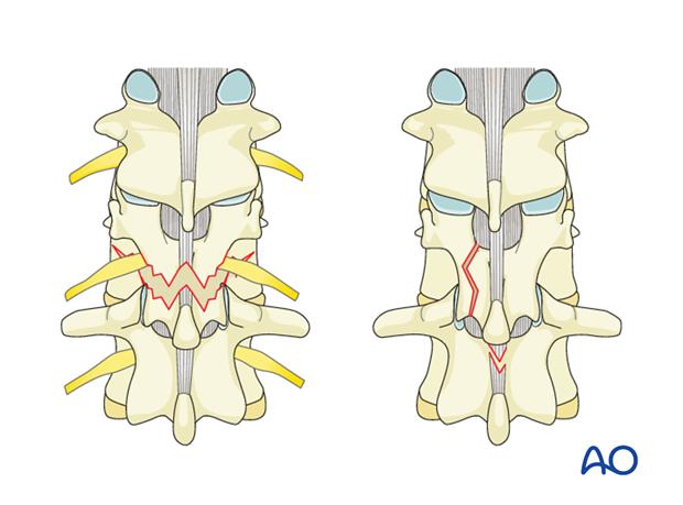
Pitfall: type C injuries
In patients with type C translational injuries, defects in the lamina and midline ligamentum flavum are common. Care should be taken during exposure. Also in type C injuries, it is better to expose the spine on one side first and place a temporary rod over the pedicle screws. Bilateral exposure in these instances can potentially destabilize the spine making it prone for further neural injury.
3. Closure
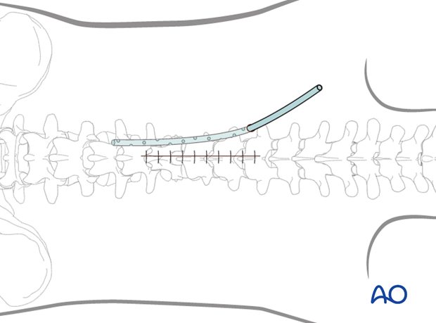
Drains are usually inserted via a separate stab incision.
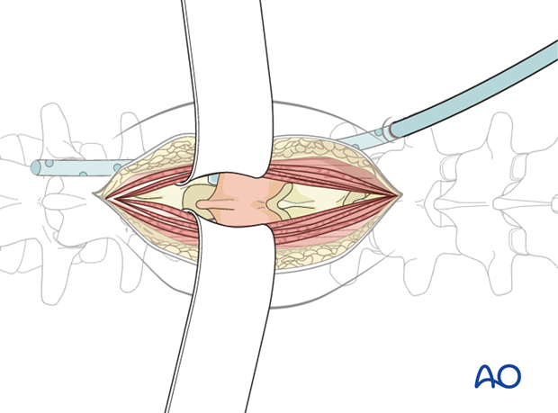
Once the surgical fixation and decompression have been performed, tight closure of the muscle and fascial layer is performed with continuous or interrupted sutures.
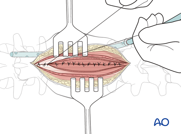
The subcutaneous layers and skin are sutured.
