ORIF - Lag screw fixation
1. Introduction
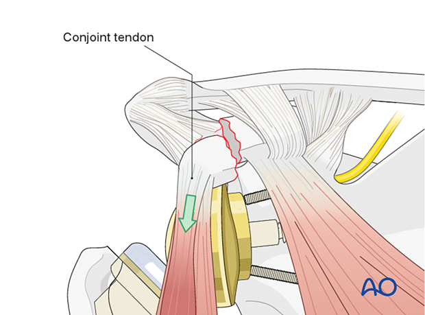
With a reverse prosthesis, there may be more tension on the conjoint tendon due to lengthening of the arm. This may result in an avulsion of the coracoid.
Coracoid fractures may occur:
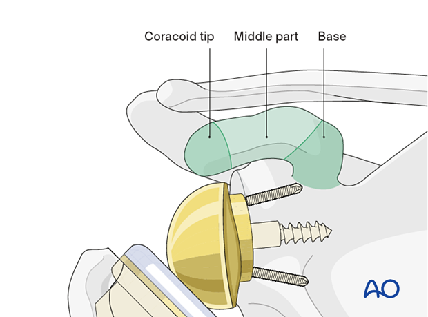
- Distal to the suspensory coracoclavicular ligaments (body and tip)
- Proximal to the suspensory coracoclavicular ligaments (base)
These fractures may occur either perioperatively, or after some delay.
Any disrupted ligaments are treated as described in the AO Surgery Reference scapula module section on the lateral scapular suspension system (LSSS).
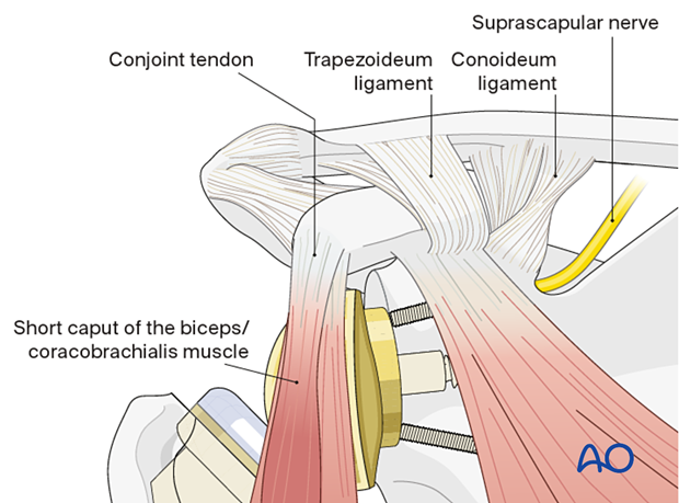
The coracoid projects anteriorly and inferiorly with a curved undersurface. The coracoid is divided into three parts: the anterior part (tip) bends forwards and downwards, the middle part is flat, and the posterior part runs to the base. This anatomical configuration must be kept in mind when screw fixation is used.
2. Patient preparation
The standard patient position is the beach chair position with inclination of about 30°. An arm holder may be helpful but is not essential.
Intraoperative fluoroscopy can be helpful.
Patient positioning should be discussed with the anesthetist.

3. Approach
A superiorly extended deltopectoral approach may be used.
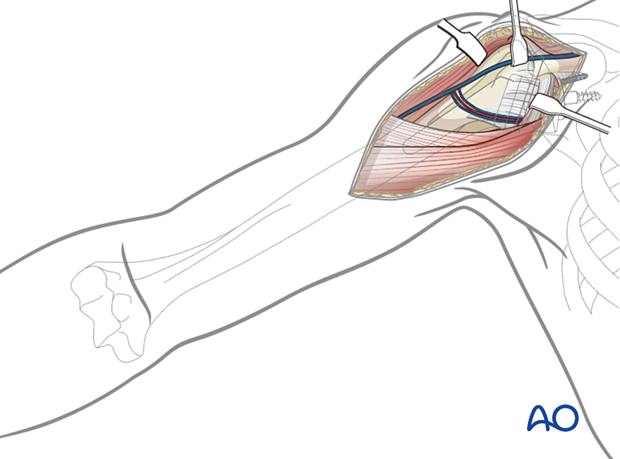
If the fracture line is lateral/distal to the coracoacromial ligaments, reduction may be made difficult by the pull of the conjoint tendon.
In this case the approach is extended superiorly to allow for the placement of a reduction clamp.
4. Reduction and fixation
The fracture is reduced with a clamp.
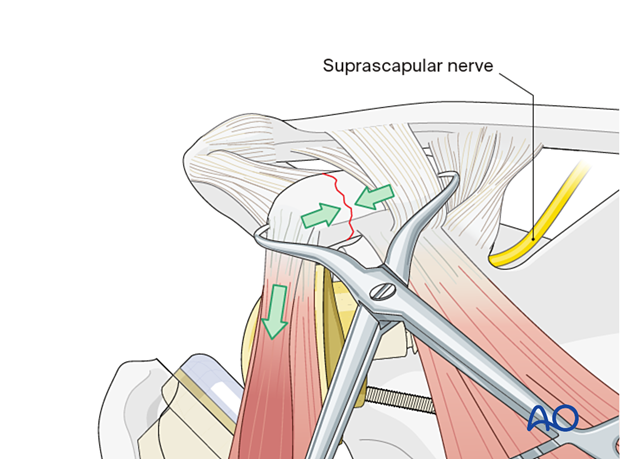
Once reduced, a K-wire is inserted for temporary fixation.
Care must be taken that the K-wire (and screw) do not enter the suprascapular notch as this may damage the suprascapular nerve (which controls the supra- and infraspinatus muscles).
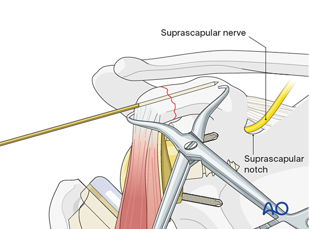
Check the reduction and temporary fixation with an image intensifier.

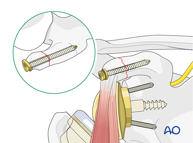
Insert the lag screw or positioning screw over the K-wire. Then remove the K-wire and clamp.
Options for fixation include:
- Standard lag screw
- Cannulated lag screw
- Cannulated headless compression screw
Use of a washer, as shown, may be indicated in osteopenic bone.
A standard lag screw with washer is shown here.
More information about the use of lag screws can be found here.
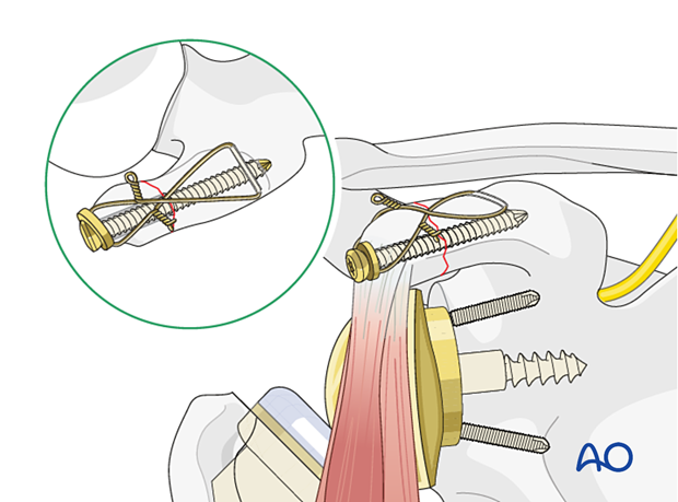
Option: cerclage to increase stability of the construct
A hole is drilled through the bone at the base of the coracoid. Make sure that this does not interfere with the screw.
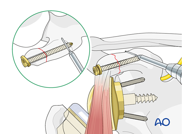
A cerclage wire or a strong nonresorbable suture is inserted and secured in a figure-of-eight fashion.
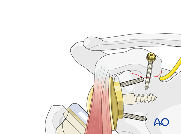
Base fractures
Base fractures are fixed in a similar way to tip fractures; however, the position of the screw is more posterior and almost vertical.
5. Aftercare
Postoperative phases
The aftercare can be divided into four phases of healing:
- Inflammatory phase (week 1–3)
- Early repair phase (week 4–6)
- Late repair and early tissue remodeling phase (week 7–12)
- Remodeling and reintegration phase (week 13 onwards)
Full details on each phase can be found here.


