Open reduction; screw and plate fixation
1. Goals
The main goals of treatment of these fractures are:
- Anatomical reduction and restoration of the joint surface
- Avoidance of joint stiffness by early motion
- Uncomplicated healing
- Avoidance of malunion or nonunion
2. Preoperative planning
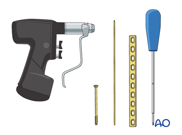
Instruments and implants
- Standard orthopedic instrument set
- Appropriate plates, according to the size of the child (2.7-3.5 standard plates, recon plates, or anatomically preshaped plates)
- Lag screws (if possible cannulated)
- K-wires
Anesthesia and positioning
General anesthesia is recommended and a sterile tourniquet should be available.
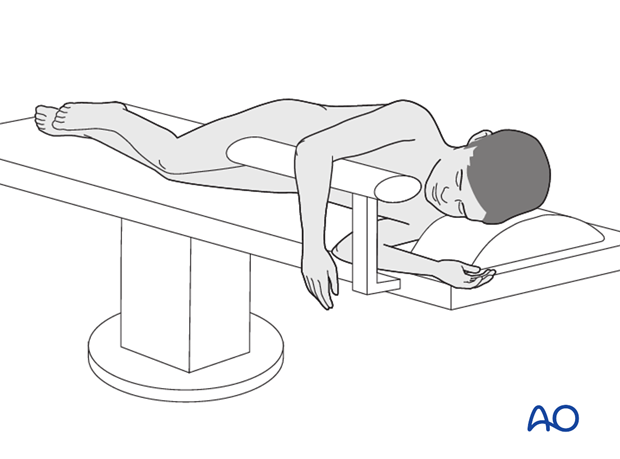
The patient should be placed lateral with the injured arm placed over a padded arm roll or a gutter support.
3. Approach
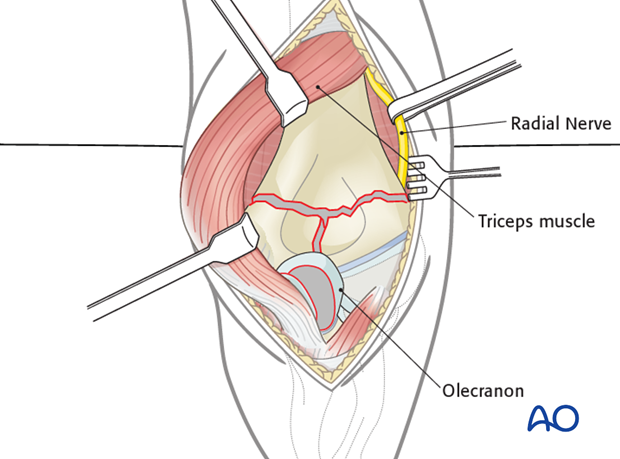
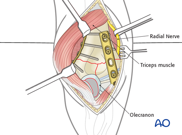
A posterior approach is used. The triceps may be split or reflected laterally and medially. Exposure may be enhanced with an olecranon osteotomy when necessary.
Note: If it has been necessary to perform an olecranon osteotomy, the repair by cerclage compression wiring is as described in the adult surgery reference for fracture types 13-C.
This form of fixation was referred to as “Tension band wiring”. We now prefer the term “Cerclage compression wiring” because the tension band mechanism cannot be applied consistently to each component of the fracture fixation. An explanation of the limits of the Tension band mechanism/principle can be found here.
Cleaning of the fracture site
The fracture is cleaned by removing blood clots, loose pieces of bone, and any interposed tissue.
4. Reconstruction of the articular surface
Condylar reassembly
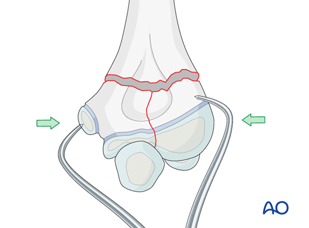
The articular fragments are reduced using pointed reduction forceps, a towel clamp, or with the help of two K-wires.
An anatomical reconstruction of the joint surface is mandatory.
Definitive interfragmentary fixation
A lag screw is used to obtain interfragmentary compression of the articular fracture plane. A partially or fully threaded screw can be used. If a fully threaded screw is used, it is necessary to overdrill the near fragment.
To achieve an optimal mechanical outcome, the physis can be ignored as these children are approaching skeletal maturity.
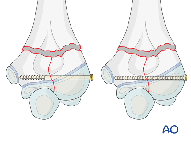
If cannulated lag screws are available, the two reduced fragments are temporarily fixed with a K-wire.
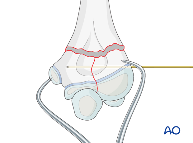
The guide wire for the cannulated screw is inserted through the center of the capitellum, parallel to the joint surface.
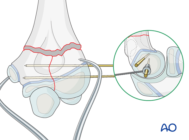
An appropriate screw, depending on the fragment size (3.0, 3.5, or 4.0 mm), is inserted and both wires are removed, unless the first K-wire is required for additional stability.
Note: Once the condylar mass has been reconstructed, the injury has been converted to a simple supracondylar fracture type.
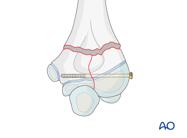
5. Condylar reattachment with biplanar plates
The reconstituted condylar mass is reduced to the metaphysis and crossed K-wires are used for preliminary fixation.
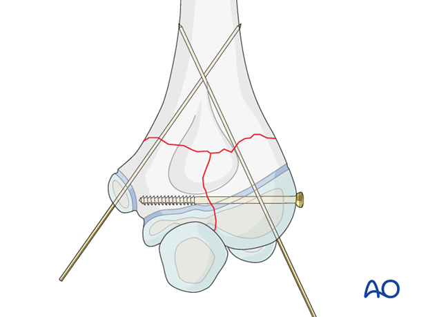
Plate preparation
The plates must be carefully contoured using appropriate malleable templates. For ease of contouring, reconstruction plates are used.
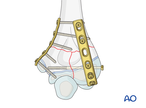
The lateral column plate is placed posteriorly and the medial column plate medially.
Note: Anatomically precontoured plates can be very useful if the bone size matches the plate shape.
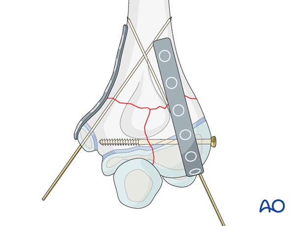
Definitive fixation
A 3.5 mm reconstruction plate is placed posteriorly. It may curve around the capitellum which is nonarticular posteriorly.
In a distal fracture, the reconstruction plate can be bent all the way to the edge of the capitellar articular surface. It will not interfere with the radial head during extension of the joint. The more bone that is covered by the plate, the greater the stability.
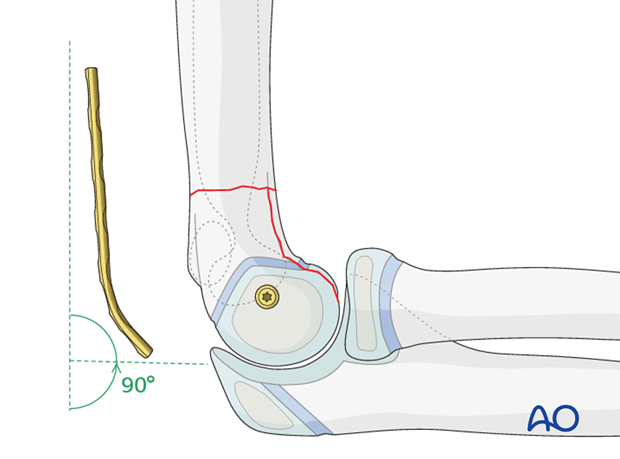
Application of the lateral plate
One or two screws are inserted through the distal portion of the plate and the most proximal screw is then inserted eccentrically as a load screw. As this pulls the plate proximally, the metaphyseal fracture plane is compressed.
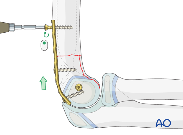
Screw insertion
The plate is fixed to the bone by inserting the remaining screws in neutral mode.
Medial plate
Another plate is applied onto the crest of the medial supracondylar ridge; its orientation at a right angle to the plane of the lateral plate increases stability.
It is recommended that the distal screw be angled distally to purchase in the trochlea.
Note: Confirm satisfactory fixation by imaging at the conclusion of the operation.
6. Aftercare
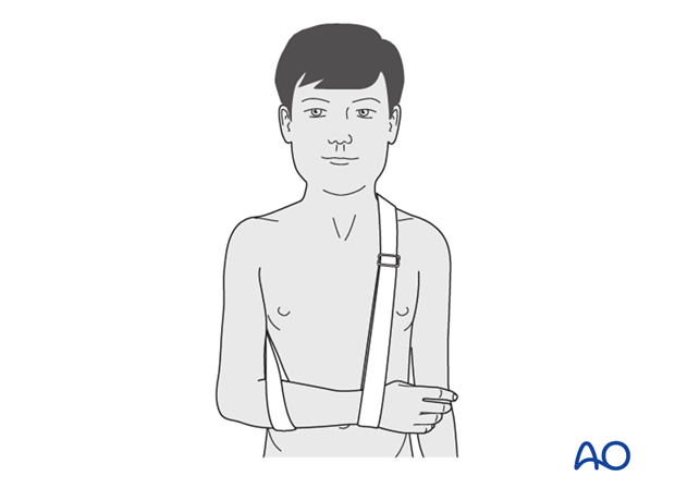
Plating provides stable fixation and rarely requires prolonged external immobilization. A soft dressing or backslab is used for comfort for no more than 10 days.
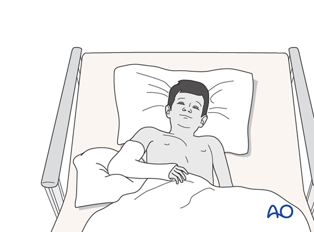
If the child remains for some hours/days in bed, the elbow should be elevated on pillows to reduce swelling and pain.
See also the additional material on postoperative infection and compartment syndrome.
The postoperative protocol is as follows:
- Discharge from hospital according to local practice (1-3 days)
- First clinical follow-up for cast removal is undertaken at approximately 7-10 days postoperatively. Gentle active assisted elbow range of motion exercises are taught
- At 4-5 weeks postoperatively, the child is reviewed clinically and radiologically
- In most cases, at this x-ray control, the fracture is usually consolidated and stable
Removal of plates
In the older child, the plates may not need removal unless they are symptomatic. In any case plate removal should not be undertaken before full consolidation and modelling of the fracture is seen radiologically.
Plate removal is performed under general anesthesia and may require an overnight hospital stay.


