Closed indirect reduction, K-wire fixation
1. Goals
The main goals for treatment of these fractures are:
- Uncomplicated healing
- No secondary displacement
- Avoiding overgrowth due to unstable fixation
- Avoiding secondary complications, such as nonunion, leading to additional problems (eg, ulnar nerve irritation)
It is important to be aware that K-wire fixation gives less stability than screw fixation.
2. Introduction
Suitable fracture type
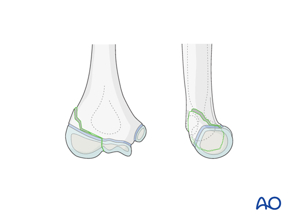
13-E/4.1L with no more than 2 mm displacement in the metaphyseal area and stable in the joint ("hanging fracture").
General considerations
Normally, all displaced intraarticular fractures (which in children always are epiphyseal fractures) require absolute anatomical reduction, which can only be performed by an open method. However, for "hanging fractures" minimally invasive treatment can be attempted.
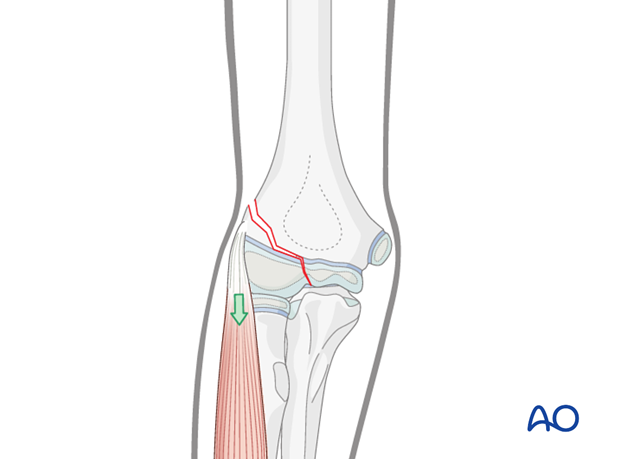
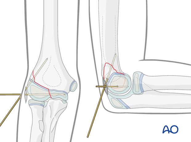
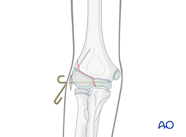
The problem of any nonoperative treatment of this fracture is that the fracture can be completed and displaced by extensor muscle tension, thereby becoming unstable (as illustrated). It is therefore recommended that the fracture be fixed by insertion of two divergent K-wires.
3. Preparation
Instruments and implants
As this is a minimally invasive surgical technique, only a few instruments are necessary.
1.2 mm, or better 1.6 mm, K-wires should be used.
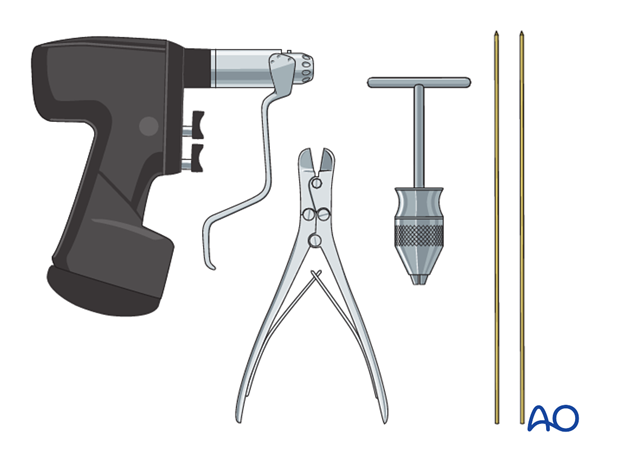
The following equipment is needed:
- K-wires of appropriate size
- Drill, preferably oscillating to avoid thermal injury, or T-handle
- Wire cutting instruments
- Image intensifier
- If necessary, contrast medium for arthrography
Anesthesia and positioning
General anesthesia is recommended and a sterile tourniquet should be available. Muscle relaxation is not necessary.
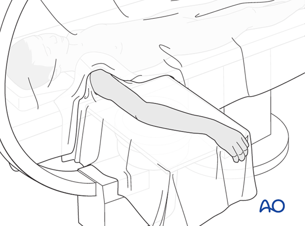
The patient is placed supine with the arm draped up to the shoulder.
4. Reduction and fixation
Planning of the entry point
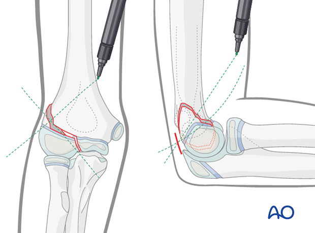
In the AP view, a line is drawn from the most prominent lateral part of the capitellum and perpendicular to the fracture line.
The arm is positioned for a true lateral view and a line is drawn from the posterior metaphyseal fragment to the anterior cortex (as shown in the illustration), perpendicular to the fracture line.
The crossing point of these two lines on the skin is the entry point for the K-wire.
Skin incision and approach to the bone
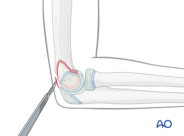
A small (5 mm) skin incision is made using a pointed knife.
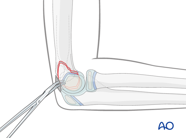
The wound is deepened by blunt dissection using a small artery forceps, down to bone contact.
Insertion of first K-wire
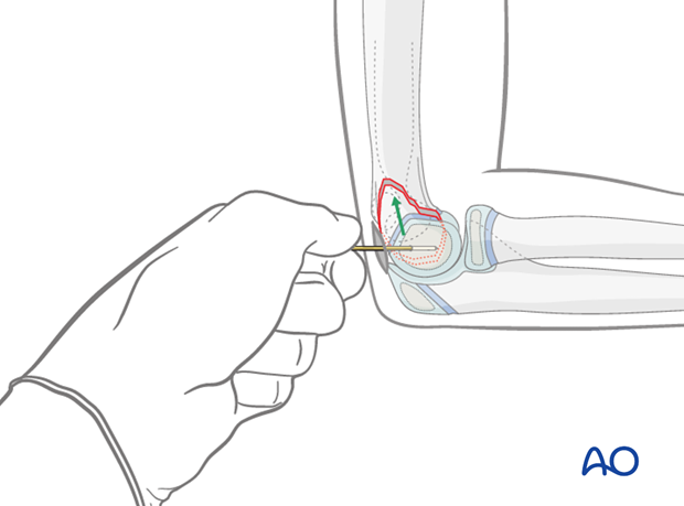
A 1.2 or 1.6 mm K-wire is inserted parallel to the joint surface by hand until it has good bony contact.
The K-wire is pressed 1-2 mm inside the cartilage of the capitellum.
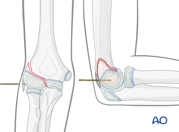
The direction of the K-wire is checked in AP and lateral views.
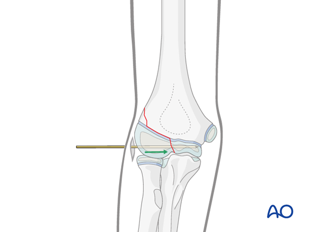
Once the position is correct, the K-wire is used as a joystick to achieve reduction and then advanced into the capitellum.
If the arthrography reveals that the joint surface is aligned, the K-wire is advanced to the medial side.
Note: Care must be taken not to over penetrate and risk compromise of the ulnar nerve.
Insertion of second K-wire
A second K-wire is inserted posterior to the first K-wire and advanced proximally through the lateral column of the humerus as far as the anterior cortex.
Trimming of K-wires
Whether the K-wires are left outside the skin or bent and buried beneath the skin depends on the surgeon's preference.
It is possible to bend the wires under the skin and hammer the bent wires further into the bone. This provides slightly more stability and prevents pin-track infection. However, a second general anesthesia (or local anesthesia in older children) is required for wire removal.
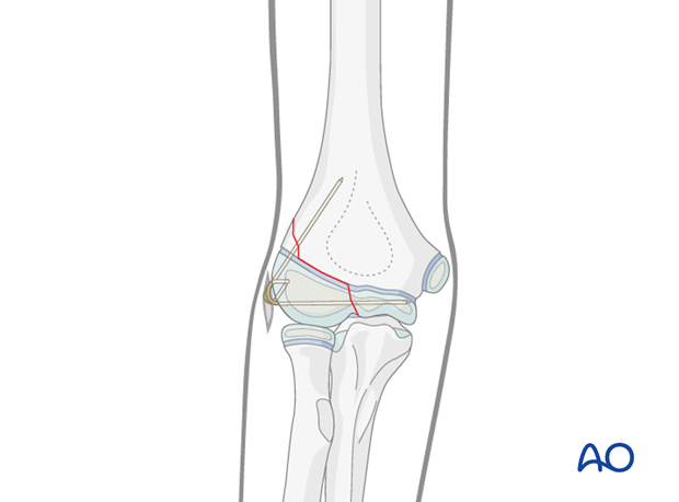
K-wires may be left protruding, but there is a risk of pin-track infection. The advantage is that the K-wires can be removed without anesthesia.
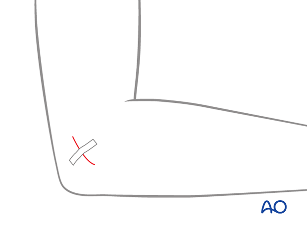
The small incision is closed with one suture or a strip.
5. Aftercare
Note: K-wire fixation alone confers minimal stability. Therefore, additional cast immobilization is required to reduce the risk of secondary displacement. Recommended immobilization for K-wire fixation is posterior and anterior long arm splints.
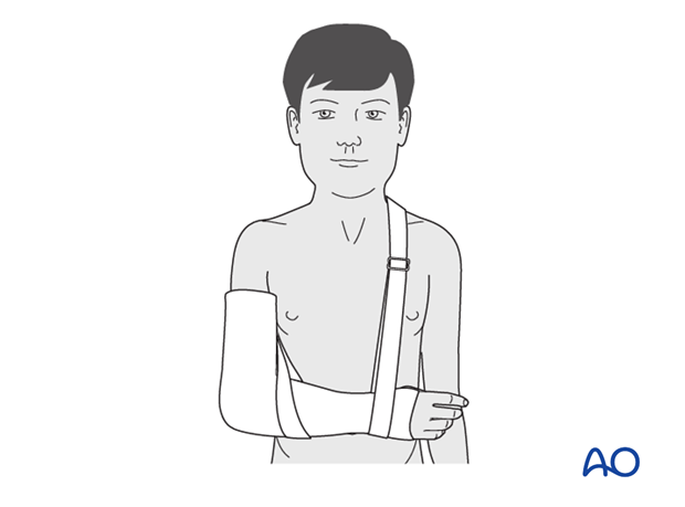
Note: In any case of elbow immobilization by plaster cast, careful observation of the neurovascular situation is essential both in the hospital and at home.
See also the additional material on postoperative infection.
It is important to provide parents with the following additional information:
- The warning signs of compartment syndrome, circulatory problems and neurological deterioration
- Hospital telephone number
- Information brochure
If the child remains for some hours/days in bed, the elbow should be elevated on pillows to reduce swelling and pain.
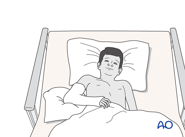
The postoperative protocol is as follows:
- Discharge from hospital according to local practice (1-3 days)
- First clinical and radiological follow up, depending on the age of the child, is undertaken 4-5 weeks postoperatively out of the cast
Note: For complete or incomplete undisplaced fractures, some surgeons recommend cast-free control x-ray after 4-5 days to verify whether the fracture remains reduced or not. If the fracture has displaced, open reduction and screw fixation is indicated. - In most cases, at 4-5 weeks, the fracture is consolidated and stable so that the cast is no longer required
- Physiotherapy is normally not indicated
K-wire removal
Protruding K-wires can be removed in the outpatient clinic 4-5 weeks postoperatively.
Buried K-wires can be removed after 2-3 months. This can be performed as a day case under light anesthesia.


