Closed reduction; K-wire fixation
1. Preparation
Timing of treatment
Active intervention is only required if, at the time of admission, the separation of the physis is still mobile.
Instruments and implants
It is recommended to use 1.0 or 1.2 mm K-wires.
The following equipment is needed:
- K-wires (1.0 or 1.2 mm)
- Drill (in case K-wires are not inserted by hand), preferably oscillating to avoid thermal injury, or a T-handle for manual insertion
- Wire cutting instruments
- If necessary, contrast medium for arthrography

As most of this epiphyseal fragment is cartilage and the wires are transphyseal, manual insertion with a T-handle is recommended.
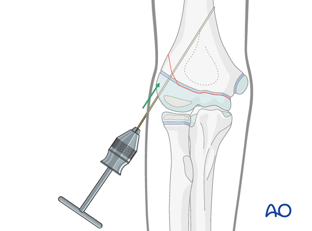
Pearl: If a T-handle is not available the K-wire can be bent into a U-shape and manually inserted (see illustration).

Anesthesia and positioning
For children, general anesthesia is always recommended. Muscle relaxation is not necessary.
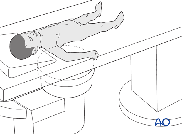
The patient is placed supine with the arm draped up to the shoulder.
2. Technique
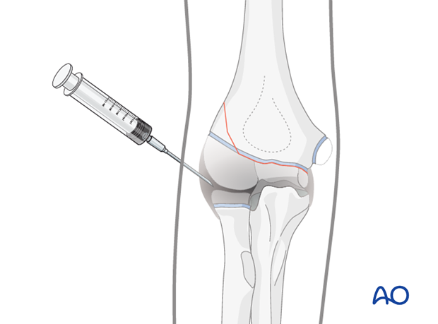
Arthrography is indicated when the fragment cannot clearly be identified.
No force is necessary to realign the epiphyseal fragment with the shaft. Forceful maneuvers could damage the physis.
Insertion of first K-wire
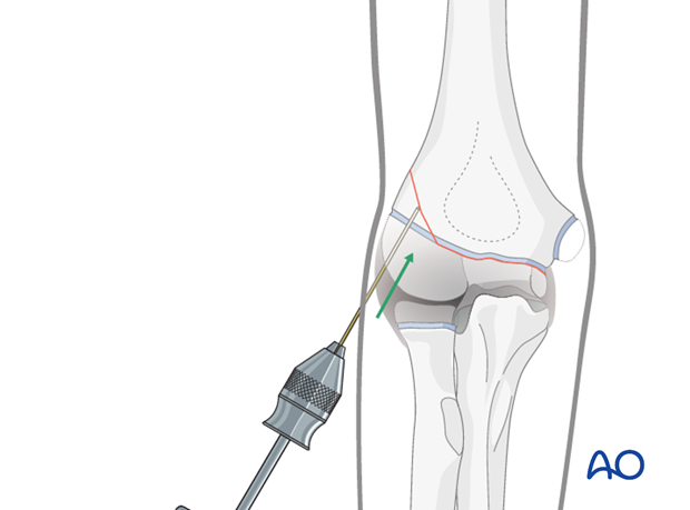
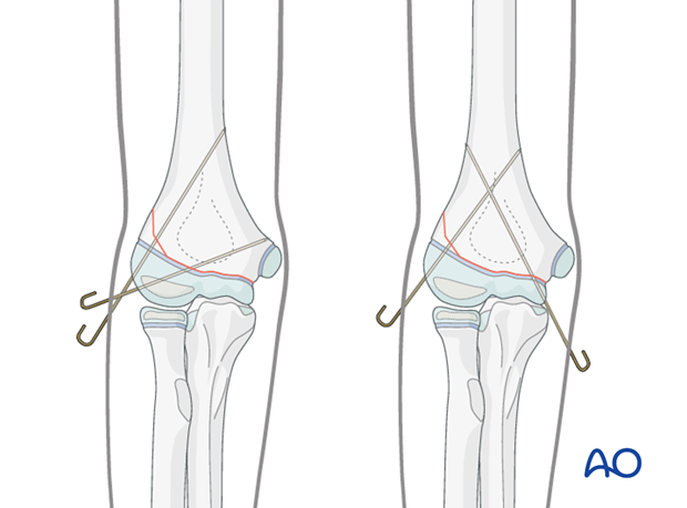
The first K-wire is inserted through a small lateral stab incision into the fragment from distal to proximal.
Before passing the fracture line, the reduction of the fragment to the metaphysis is checked using image intensification. The K-wire is then advanced into the metaphysis.
Insertion of second K-wire
A second oblique lateral K-wire is recommended.

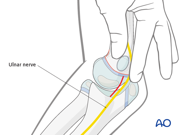
If the second K-wire is inserted from the medial side, it is recommended to visualize the insertion point with a small incision. This is to decrease the risk of damaging the ulnar nerve and ensures an appropriate insertion point.
The K-wires are bent and cut outside the skin.
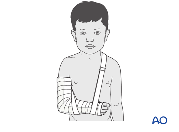
Splint application
A long-arm splint is applied with the elbow at 90°.
3. Aftercare
The postoperative protocol is as follows:
- Time in hospital according to local practice (1-3 days)
- First clinical and radiological follow up, depending on the age of the child, 2-3 weeks postoperatively out of the cast
- In most cases, at this first x-ray control, the fracture is consolidated and stable so that the cast is no longer required and the K-wires can be removed
- Physiotherapy is not usually indicated
See also the additional material on postoperative infection and compartment syndrome.


