

Intramedullary screw fixation
1. General considerations
Introduction
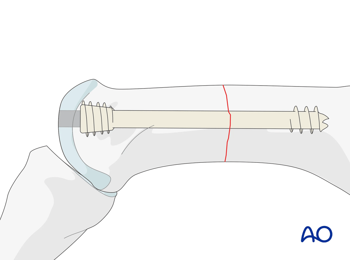
Transverse shaft and distal extraarticular fractures may be fixed with an intramedullary headless screw (3.0 or 4.0 mm).
This screw acts like an intramedullary nail.
Prerequisites for intramedullary screw fixation
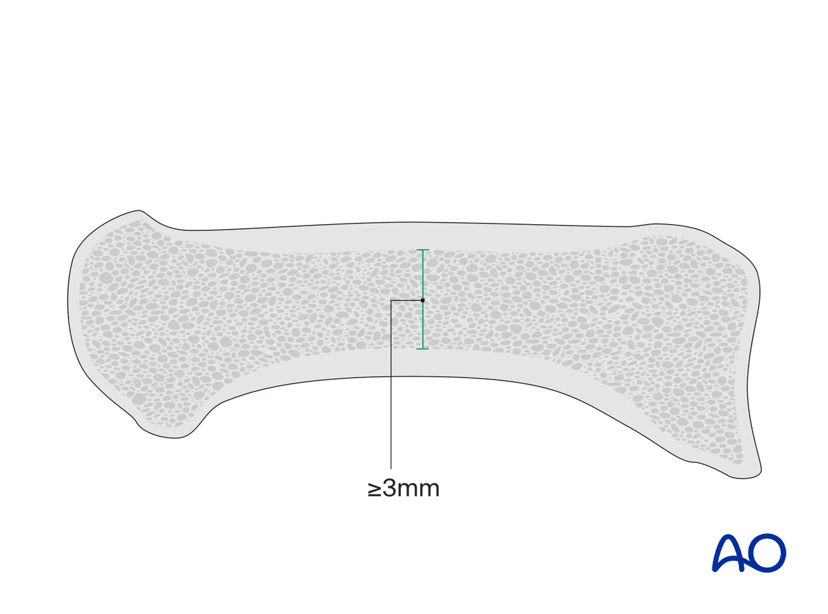
The diameter at the isthmus of the intramedullary canal should be at least 3 mm wide in AP and lateral views.
To allow for fracture compression, the length of the screw should be long enough so that the threads at the tip only engage in the far fragment.
To achieve optimal stabilization, the screw should be long enough and with a diameter filling the intramedullary canal.
The distal fragment should be long enough to allow for full insertion of the headless screw and the head thread.
2. Patient preparation
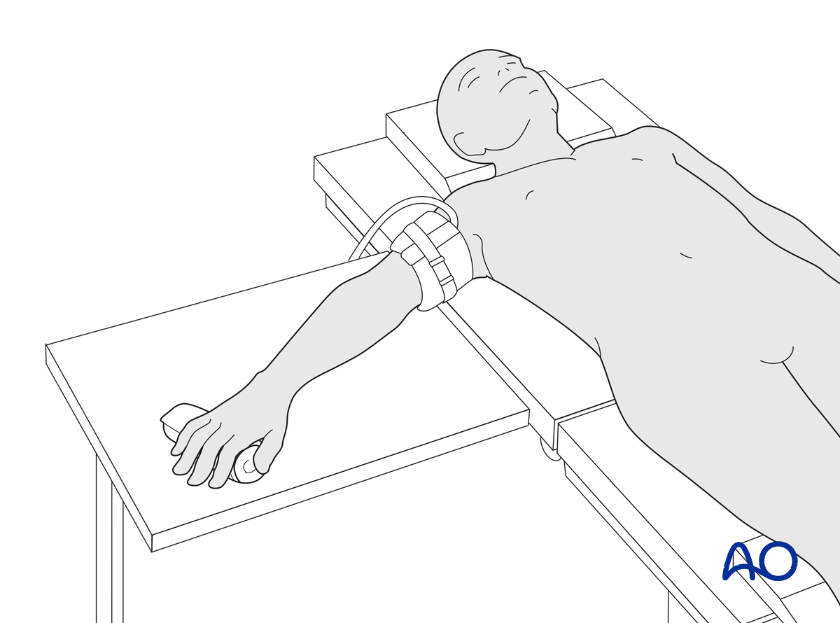
This procedure is usually performed with the patient in a supine position with the arm on a radiolucent side table.
3. Reduction
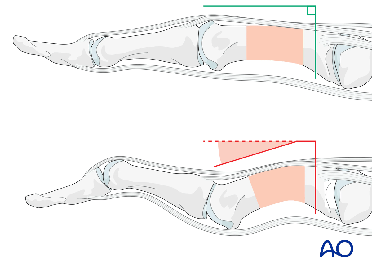
Any axial displacement should be reduced to gain full length of the metacarpal.
Closed reduction
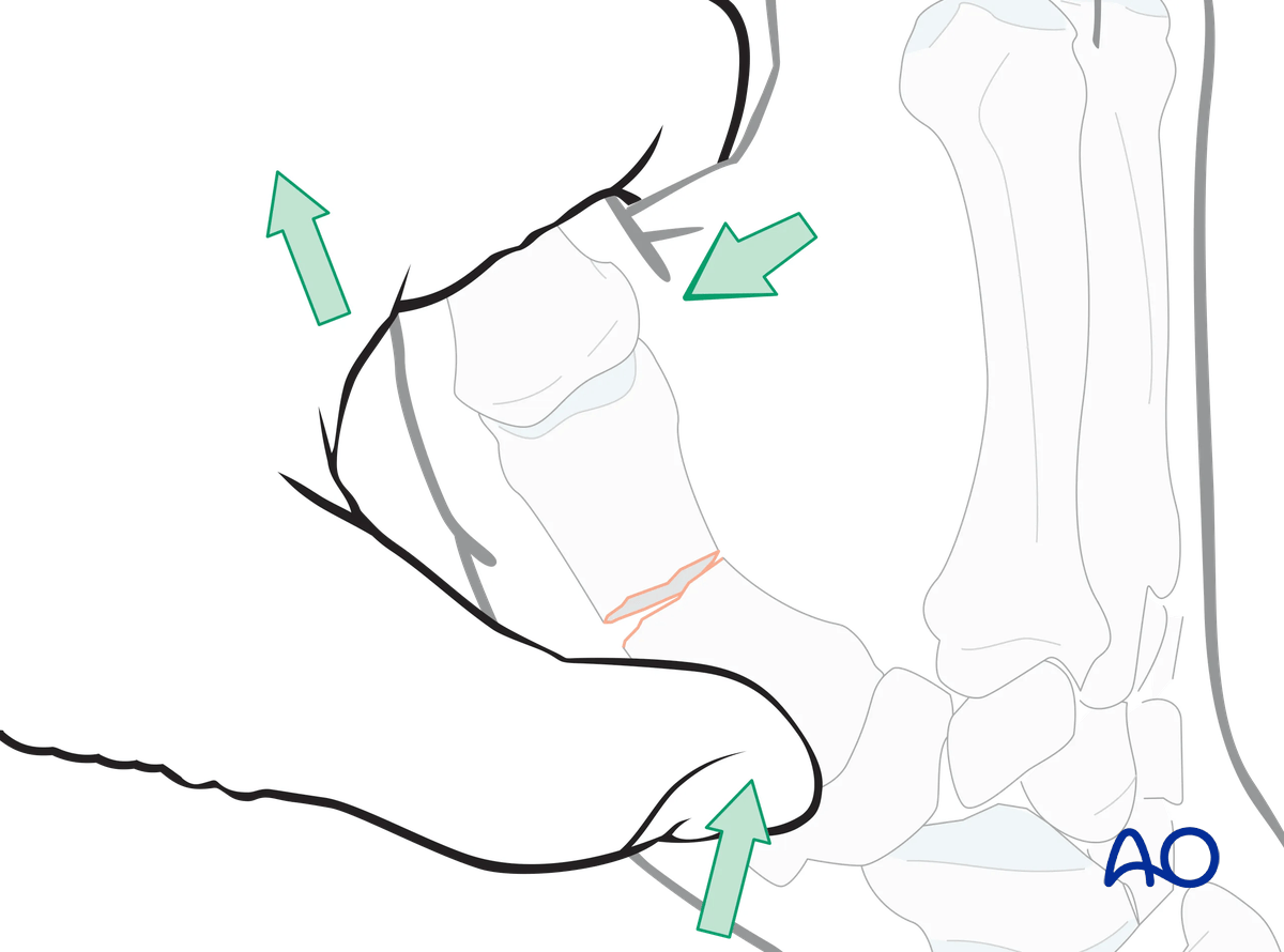
Closed reduction is achieved by applying longitudinal traction and flexing the MCP joint, then pushing the distal fragment dorsally and the proximal fragment palmarly to correct the apex dorsal angulation.
4. Checking alignment
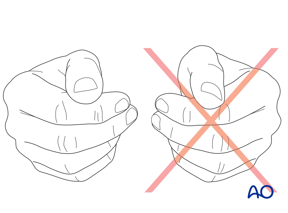
Confirm correct rotational reduction by inspecting the thumb nail plate in relation to the index finger. Compare it to the contralateral side if necessary.
5. Retrograde screw insertion
Guide wire insertion
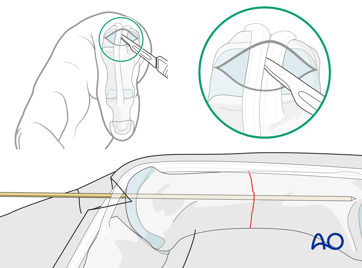
Hold the MCP joint in 90° flexion.
Incise the skin and extensor tendon and identify the dorsal part of the metacarpal head.
Insert the guide wire dorsally into the head in line with the intramedullary canal under image intensification until the tip reaches the base.
Confirm correct rotational alignment.
Screw insertion
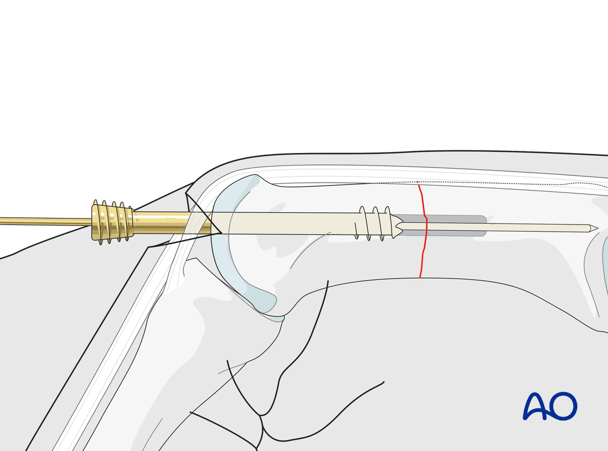
Predrill the entry point and if needed the medullary canal. This creates a track for screw insertion and reduces the risk of deviation during screw insertion.
Select a screw with the appropriate diameter and length using the depth gauge provided. The thread of the tip should engage the isthmus distal to the fracture line.
Insert the screw over the guide wire until the head is fully entered into the metacarpal and the fracture is compressed.
Confirm reduction and correct screw placement with an image intensifier.
Confirm correct rotational alignment of the finger.
6. Final assessment
Confirm correct rotational alignment by clinical examination.
Image intensification may be used to confirm anatomical reduction and correct placement of implants in two views.
7. Aftercare
Healing phases
The aftercare can be divided into four phases of healing:
- Inflammatory phase (week 1–3)
- Early repair phase (week 4–6)
- Late repair and early tissue remodeling phase (week 7–12)
- Remodeling and reintegration phase (week 13 onwards)
Full details on each healing phase can be found in the chapter Postoperative treatment of thumb injuries.
Postoperative care
Early range of motion, without load bearing, in the hand is important for optimal outcome.
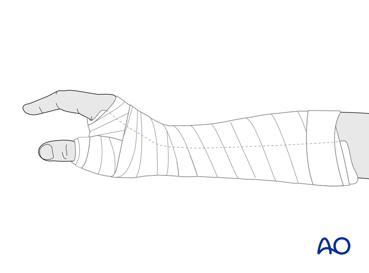
Immediately postoperatively, a temporary plaster splint is applied. This also helps with swelling and pain control.
The plaster splint can be switched to a custom-made thermoplastic splint after a week. The splint should be worn until fracture healing has been confirmed.
Hand therapy should be initiated as soon as possible.
The arm should be actively elevated to help reduce any swelling.
Mobilization
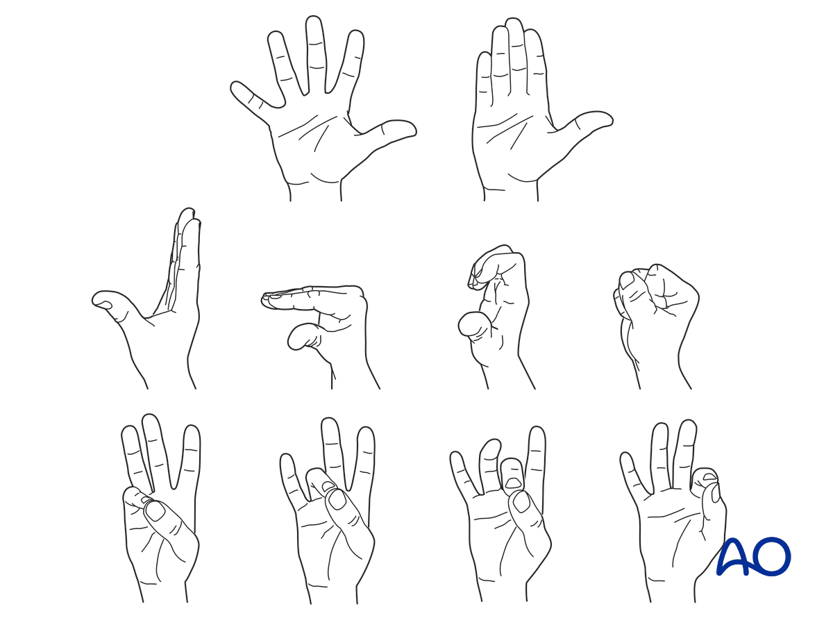
To prevent joint stiffness, the patient should be instructed to begin active motion (flexion and extension) of all nonimmobilized joints of the hand immediately after surgery.
In compliant patients with stable fixation, the splint can be removed under the supervision of a hand therapist, and early active motion exercises started after a few days.
Weight bearing should be avoided until fracture healing is confirmed.
Follow-up
The surgical wound is checked as necessary depending on soft tissue status. The wound is monitored for evidence of infection.
To allow for early joint mobilization and to prevent wound dehiscence, leave the sutures for 2 weeks.
Confirm fracture healing clinically and radiologically after about 5–6 weeks (depending on the fracture configuration).
Implant removal
The removal of an intramedullary screw is challenging and may cause damage to the articular surface.


