

Suture fixation of flexor tendon
1. General considerations
A palmar avulsion of the flexor tendon may be associated with subluxation of the interphalangeal joint.
The avulsion, with or without a small bony fragment, may be fixed with a pull-out suture.
If the IP joint is unstable, it suggests collateral ligament involvement and requires additional stabilization with a splint or transarticular K-wire. A transarticular wire should always be protected with a splint to avoid breakage.
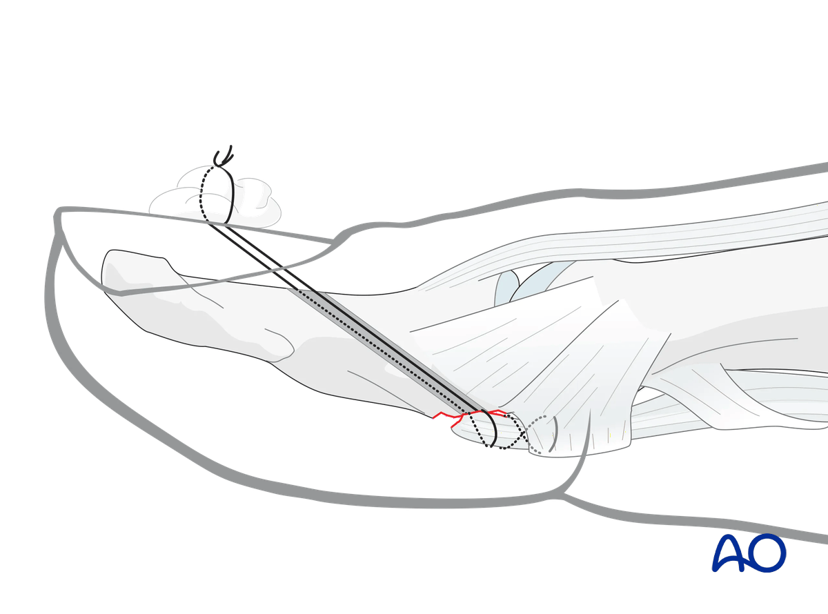
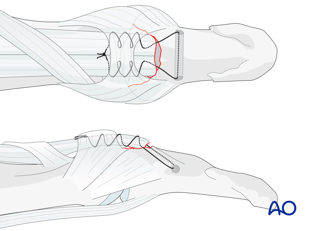
To avoid nail complications due to transungual tunneling, the transverse intraosseous loop technique may be used.
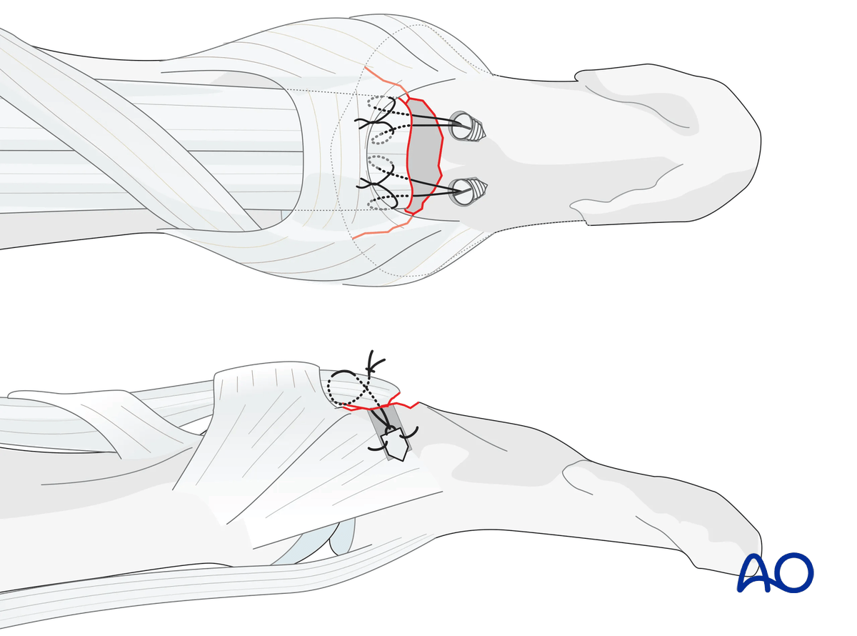
Another option to spare the nail bed is the use of suture anchors.
2. Patient preparation
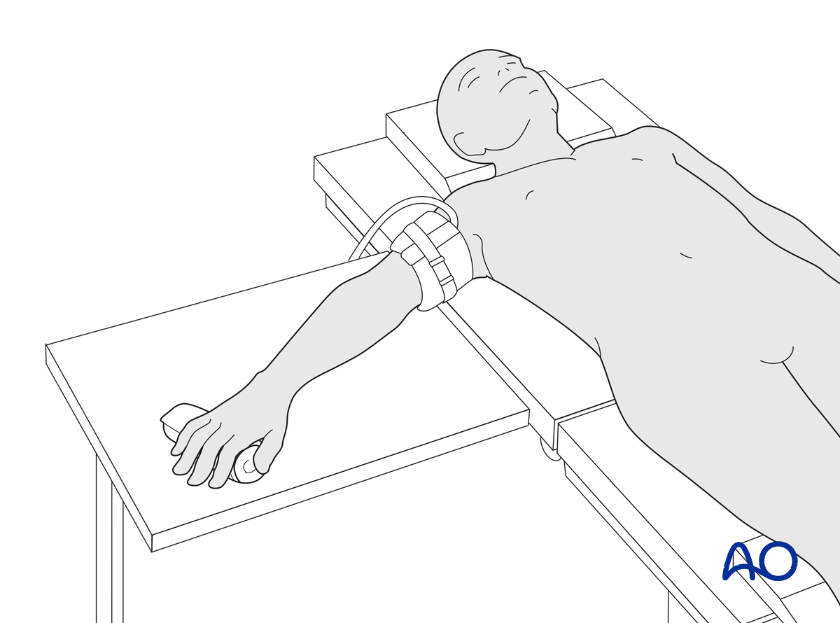
This procedure is usually performed with the patient in a supine position with the arm on a radiolucent side table.
3. Approach
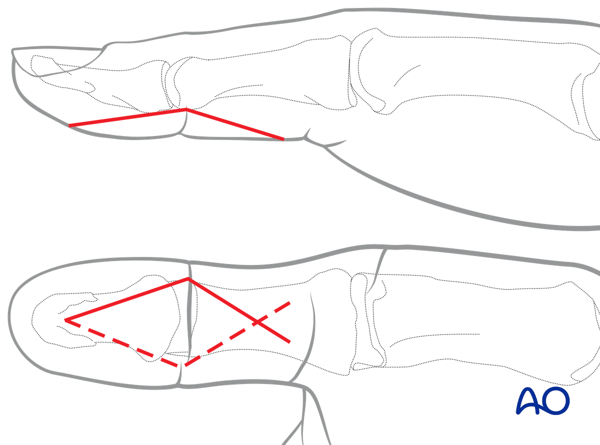
For this procedure, a palmar approach to the IP joint is typically used.
4. Reduction of dislocation
Reduction of dorsal dislocation
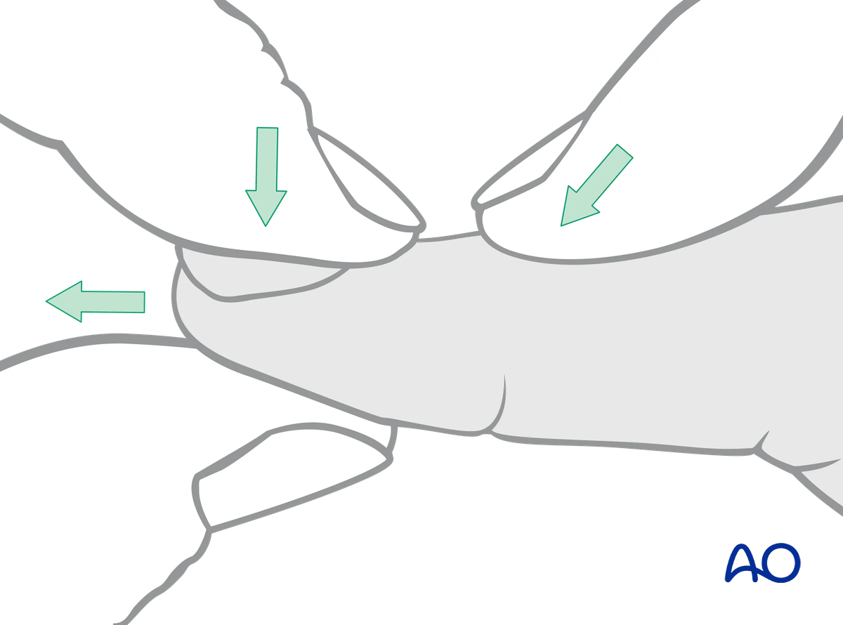
Most dorsal dislocations can be reduced by applying traction, flexion, and dorsal pressure on the base of the distal phalanx.
If the joint is stable and the flexor pollicis longus function is intact after reduction, nonoperative treatment is usually indicated.
Reduction of palmar dislocation
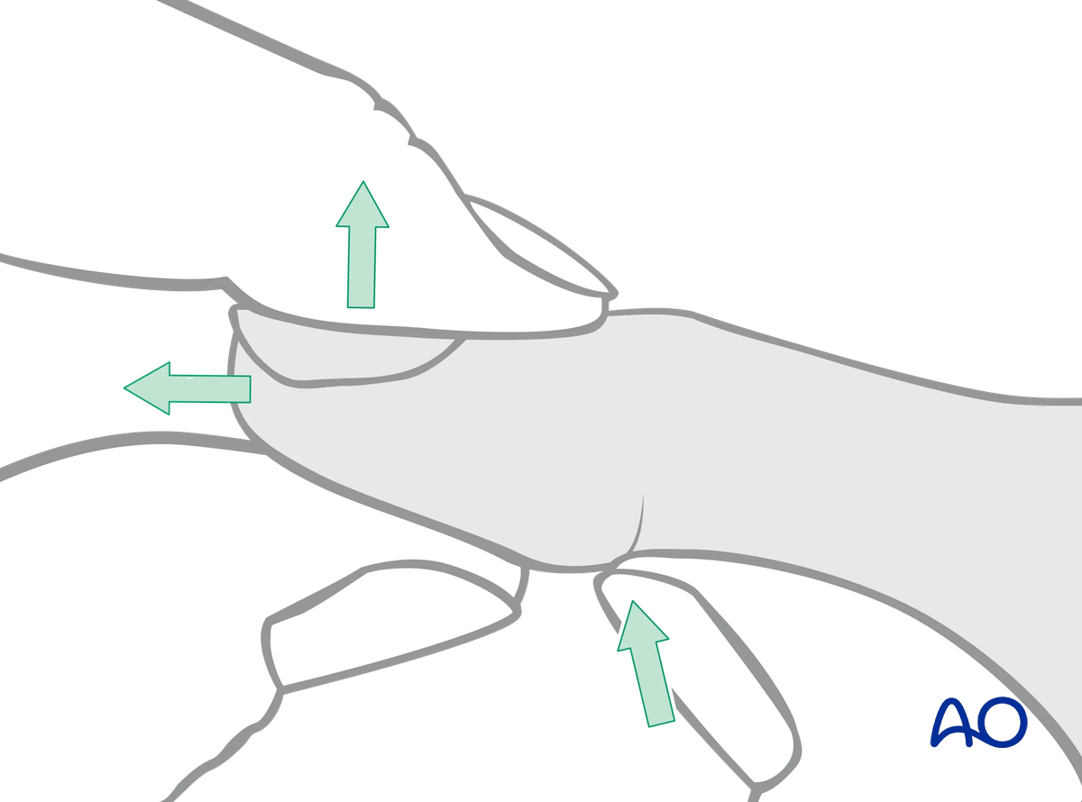
In the case of a palmar dislocation, apply traction and extension. Then apply pressure with the thumb on the palmar aspect of the base of the distal phalanx.
In palmar dislocation the terminal extensor tendon is usually disrupted.
If the joint is stable after reduction, nonoperative treatment is usually indicated.
5. Reduction of avulsion
Visualization of the joint
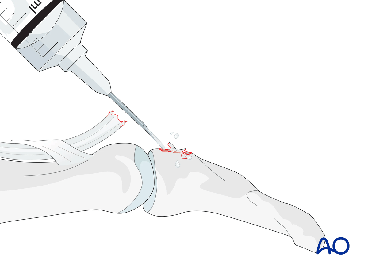
Hyperextend the distal phalanx to gain maximal visualization of the joint.
Use a syringe to clear blood clots with a jet of sterile irrigation solution.
Assess fracture geometry and look for comminution or impaction.
Often, comminution is not apparent from the x-rays and can only be determined under direct vision.
Repositioning the tendon
If the tendon has retracted to the IP joint, the zigzag incision has to be extended to the IP joint.
Use a small flexible catheter or similar tool to retract the tendon.
Use a 4.0 suture to hold the end of the flexor pollicis longus (FPL) tendon in the Mansson fashion and pass these through a suture loop. The tendon will be repositioned with a distal pull with the suture loop.
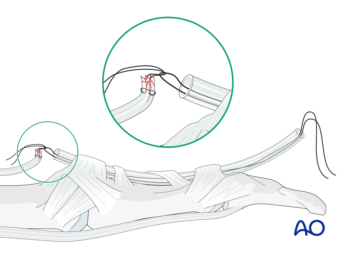
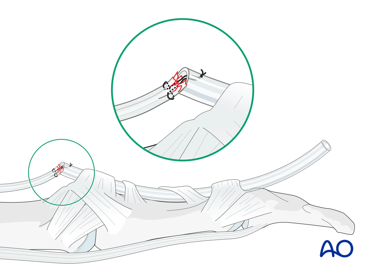
Alternatively, the FPL tendon can be sutured to the tube. By pulling it, the tendon will be repositioned.
6. Suture fixation
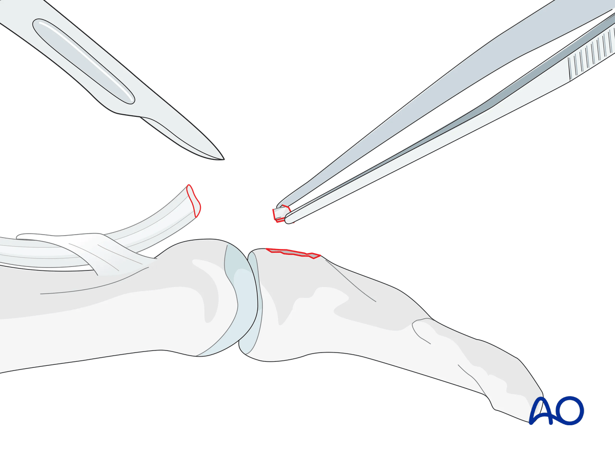
Resection of fragments
If there is comminution present, excise the comminuted bony fragments. This is also true if a flake is so small that it does not contribute to any form of reattachment.
Drilling
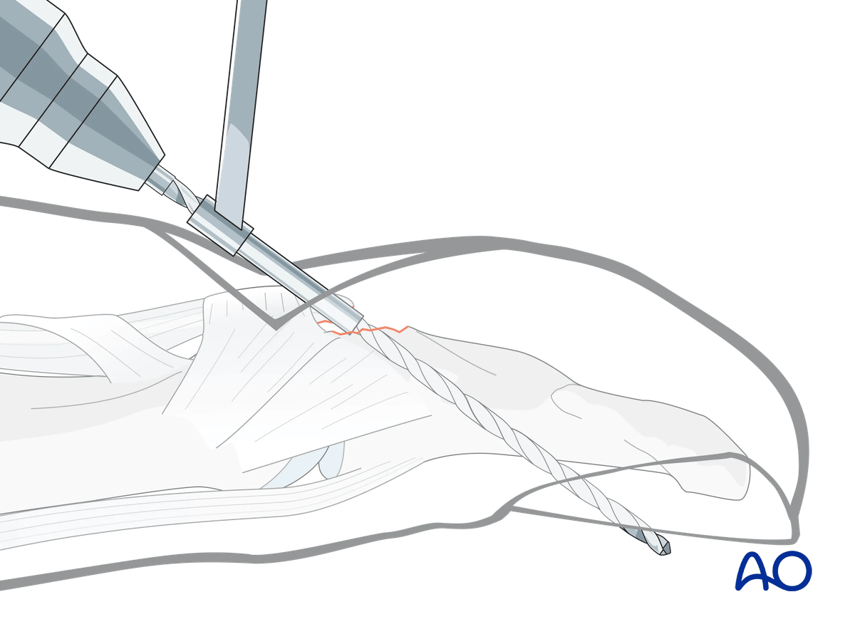
It is mandatory to use a drill guide to protect the soft tissues. Use a pillow under the hand to protect the tip of the drill bit.
Drill two parallel 1.3 mm holes aimed obliquely from proximal to distal and palmar to dorsal, exiting through the nail but avoiding the lunula (germinal matrix).
Insertion of sutures
Use 3.0 multifilament nonabsorbable sutures with double-mounted straight needles.
Interweave a suture through the detached distal end of the tendon and insert the two ends of the suture into the drill holes–both needles exit dorsally through the nail.
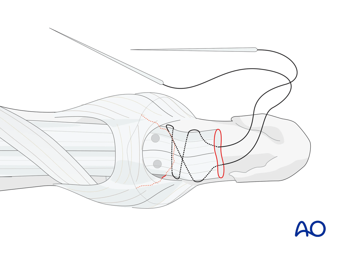
Reduction of the tendon
Gently approximate the tendon to the fracture surface by pulling the sutures in a dorsal direction.
Confirm the position under direct vision.
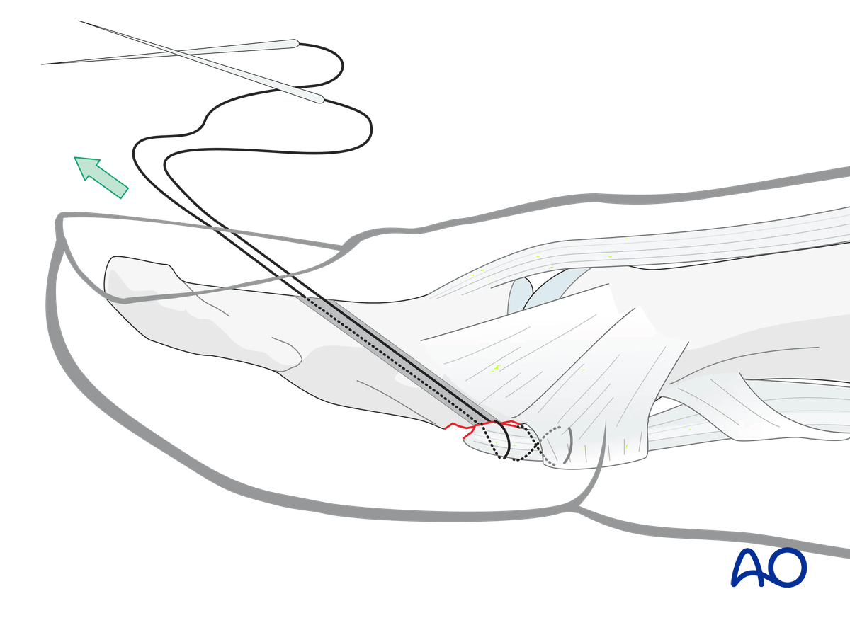
Tightening the sutures
Tighten the suture over the nail, anchoring it around a cotton piece, which also protects the nail from excessive pressure.
7. Option: transverse intraosseous loop technique
To avoid damage to the nail bed, a transosseous bone tunnel may be used for suture passing.
This tunnel is created manually using a hypodermic needle (16- or 18-gauge) in the transverse plain on the palmar aspect of the distal phalangeal base. This allows for insertion of the thread.
8. Option: suture anchors
Alternatively, to the described method, two suture anchors can be used.
This option has the advantage of sparing the nail bed and nail from injury and is a less time-consuming procedure.
Fixation of the anchors within the cancellous bone of the proximal phalanx should be assessed before passing the sutures through the bone fragment to tie them ventrally.
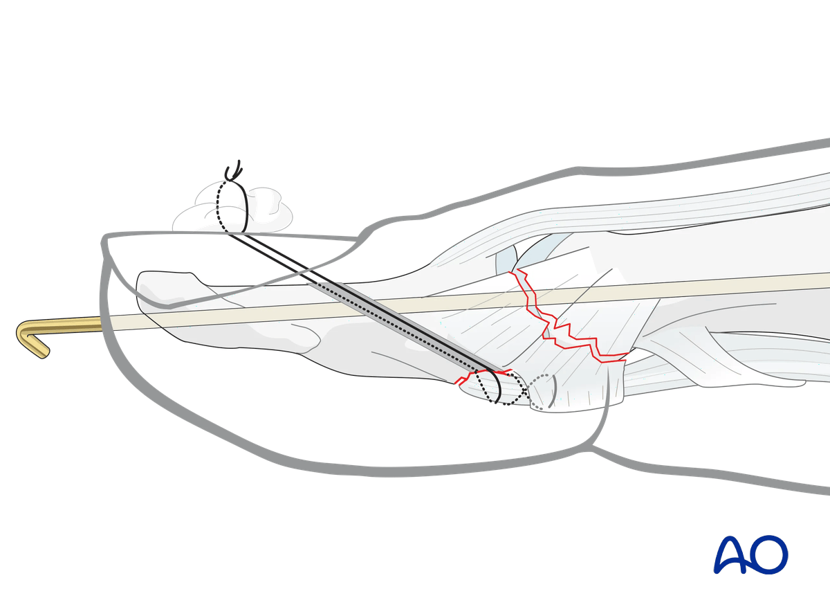
9. Joint transfixation in unstable situations
Dorsal subluxation may persist if the collateral ligaments are ruptured.
In such a case, the IP joint needs to be immobilized with a K-wire.
10. Immobilization
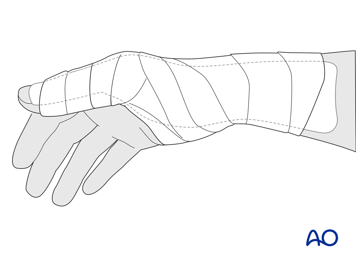
Apply a dorsal thumb spica splint with the wrist and IP joint in neutral position and MCP joint in 20° flexion for 8 weeks. This also protects a transarticular K-wire from breakage.
11. Aftercare
Healing phases
The aftercare can be divided into four phases of healing:
- Inflammatory phase (week 1–3)
- Early repair phase (week 4–6)
- Late repair and early tissue remodeling phase (week 7–12)
- Remodeling and reintegration phase (week 13 onwards)
Full details on each healing phase can be found in the chapter Postoperative treatment of thumb injuries.
Management of swelling
The splint helps with pain and edema control. The arm should be actively elevated to help reduce the swelling.
Follow-up
A wound check is performed 3–5 days after surgery. Dressings are changed to avoid the tourniquet effect of dried exudate or bleeding within the dressings.
Sutures may be removed after 10 days.
Mobilization
Hand therapy is recommended to prevent soft-tissue atrophy and joint contracture, which leads to a poor outcome, and subsequent treatment is difficult.
The fixation will be vulnerable for the first 8 weeks.
After 6 weeks, the splint can be removed during mobilization activities without weight bearing.
Splint immobilization can be completely discontinued after 8 weeks. Weight bearing exercises can be started after 12 weeks.
Suture removal
The pull-out suture may be removed after 8 weeks.


