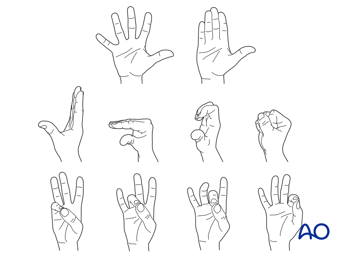
Suture fixation of extensor tendon
1. General considerations
Introduction
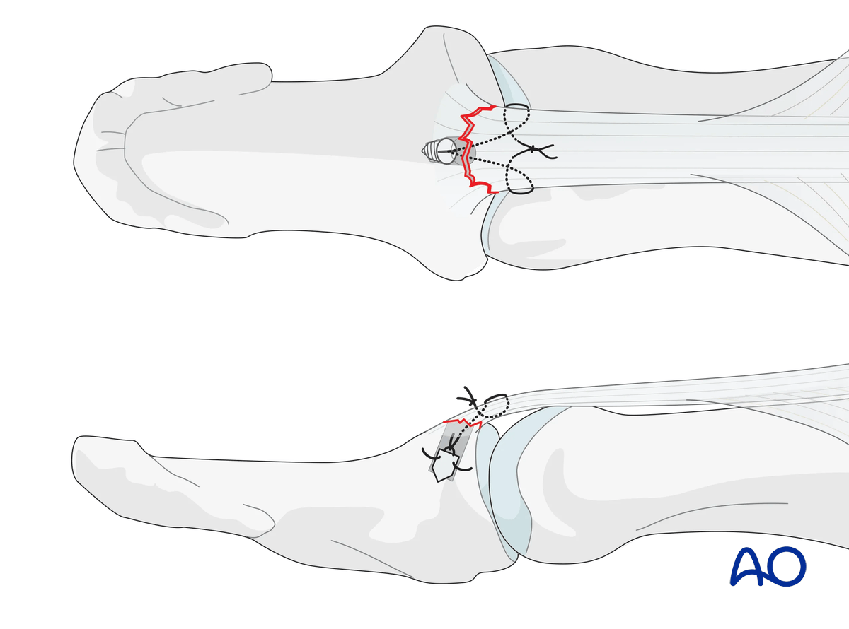
A tendinous dorsal avulsion of the distal phalangeal base caused by palmar dislocation can be fixed with a suture anchor.
Cerclage compression wiring is not recommended, as there is high risk of soft-tissue irritation and nail bed injury.
If the IP joint is unstable, it suggests collateral ligament involvement and requires additional stabilization with a splint or transarticular K-wire. A transarticular wire should always be protected with a splint to avoid breakage.
Operative treatment: caveat
Indications for surgical intervention are:
- Open fractures
- Palmar subluxation of the IP joint

Surgical management of these fractures is difficult and has many potential complications. The soft tissues are only precariously vascularized, and the fragments are very small and prone to further comminution. Healing can often be slow.
2. Surgical anatomy
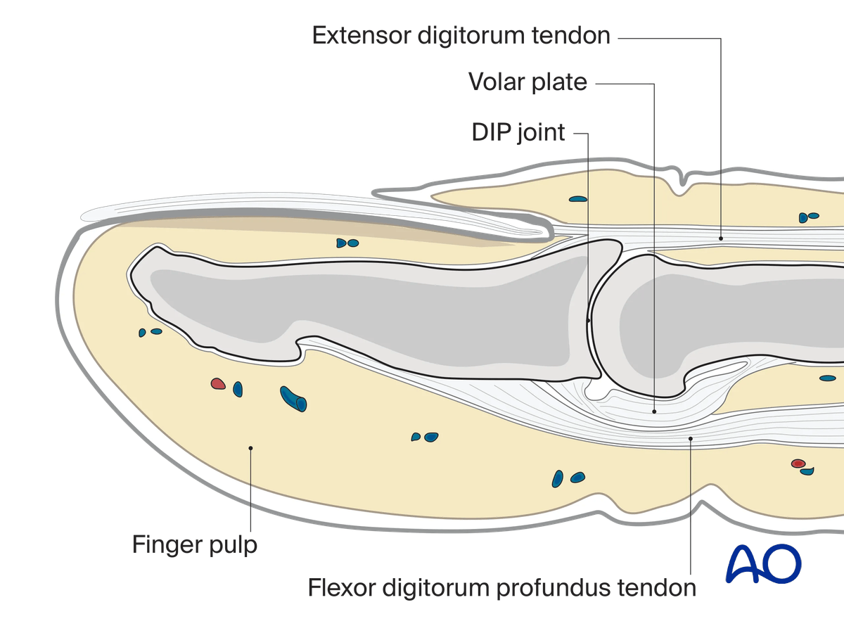
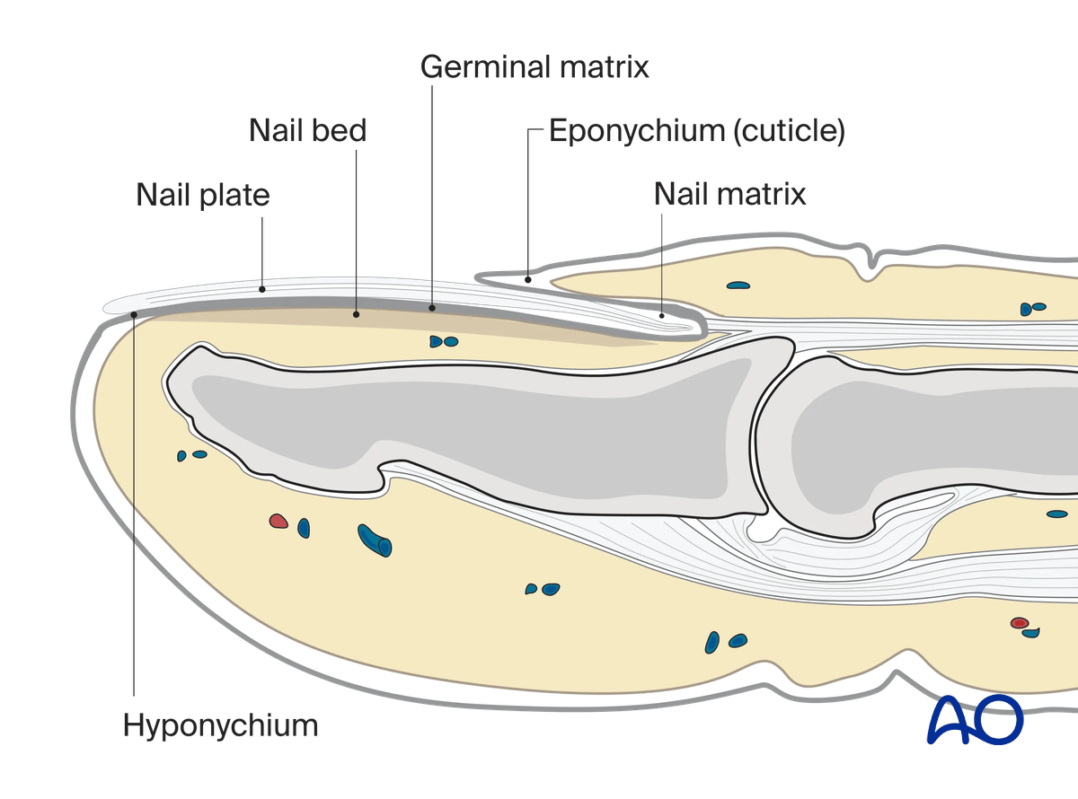
The base of the distal phalanx has a prominent dorsal crest at the insertion of the extensor tendon. The tendon is also adherent to the interphalangeal (IP) joint capsule.
On the palmar surface is the insertion of the flexor pollicis longus tendon.
The flexor tendon occupies the whole width of the base of the distal phalanx. It is made up of two different fibers. The superficial fibers attach to the lateral aspects of the phalanx. The deep fibers run centrally and attach more distal in the palmar aspect of the phalanx.
The volar plate is very flexible, allowing hyperextension of the IP joint and pulp-to-pulp pinch.
The vascularity of the extensor tendons is more precarious than that of the flexor tendons. This prolongs extensor tendon healing time.
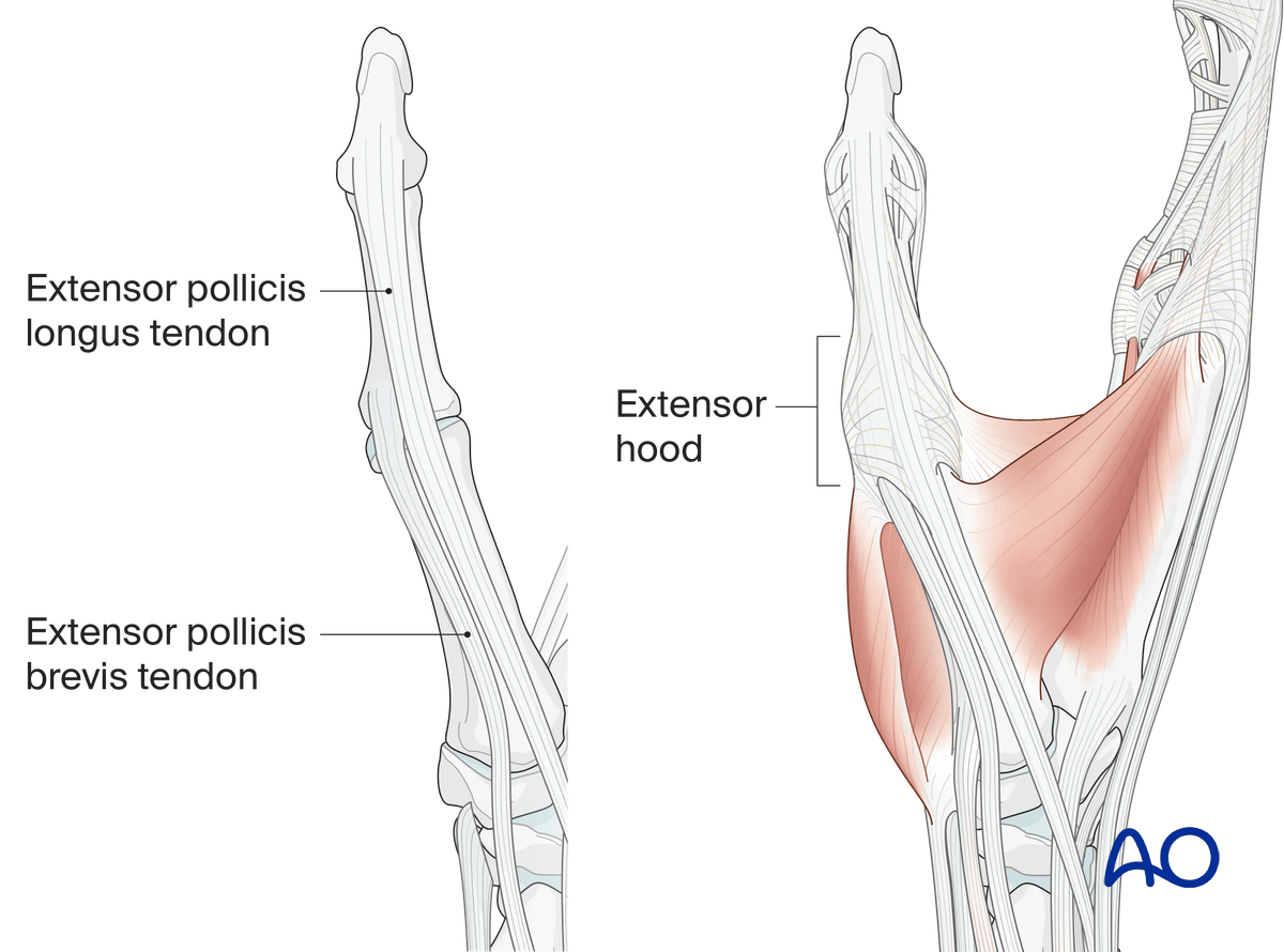
The extensor hood of the thumb consists of the radially located extensor pollicis brevis tendon (insertion: base of the proximal phalanx) and the ulnarly located extensor pollicis longus tendon (insertion at the base of the distal phalanx).
Both tendons are held in position by the sagittal bands at the level of the MCP joint and the extensor hood at the level of the proximal phalanx.
At the level of the MCP joint the abductor pollicis aponeurosis radiates into the extensor hood.
3. Patient preparation
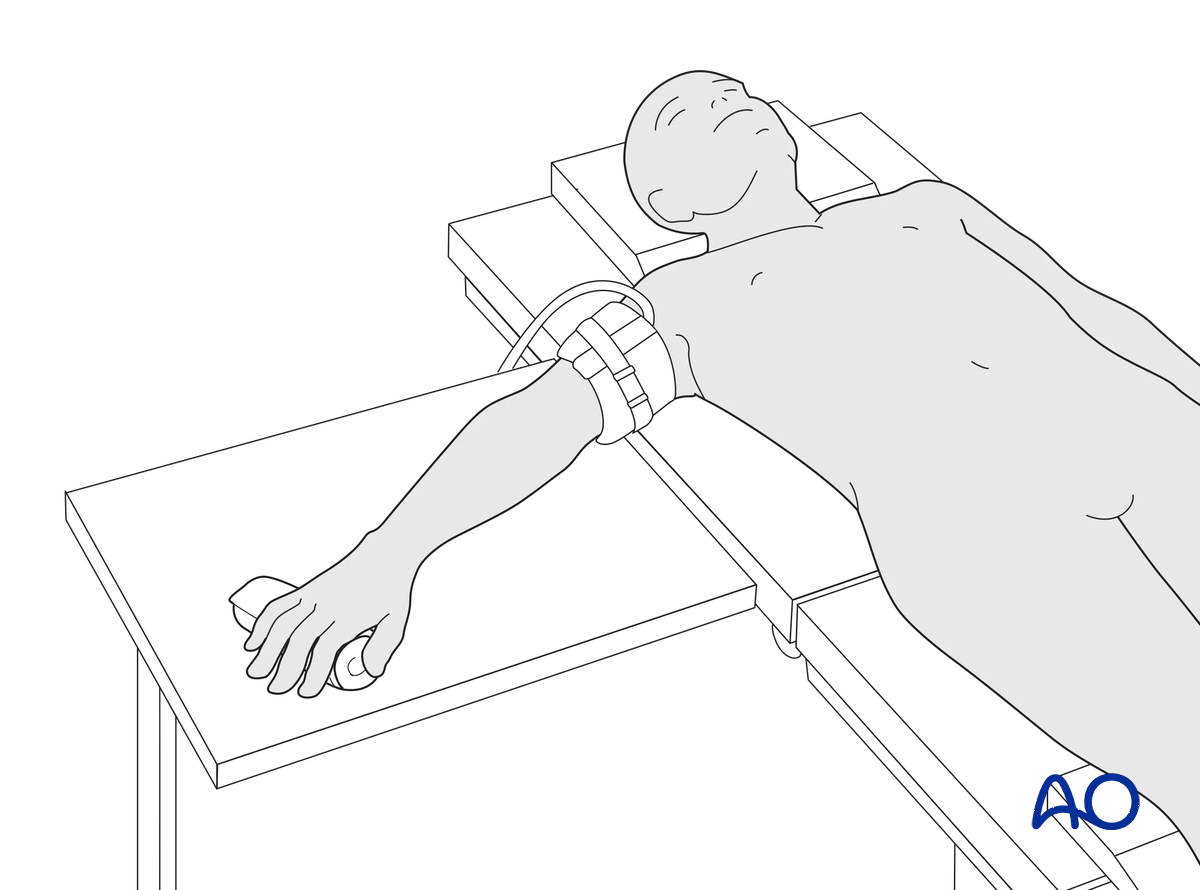
This procedure is usually performed with the patient in a supine position with the arm on a radiolucent side table.
4. Approach
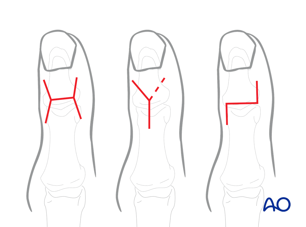
For this procedure, a dorsal approach to the IP joint is typically used.
5. Inspection
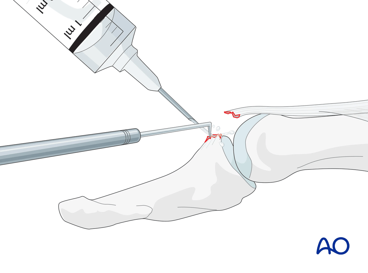
Flex the IP joint. To better visualize the rupture, use a syringe to clean out blood clots with a jet of water.
Use a dental pick to carefully free interposed tissues and remove blood clots and other debris.
6. Fixation
Use an anchor that has approximately the same width as the avulsed fragment.
Depending on the size of the fragment, two anchors can be used.
Insert the anchor according to the manufacturer’s instructions at the isometric point of insertion.
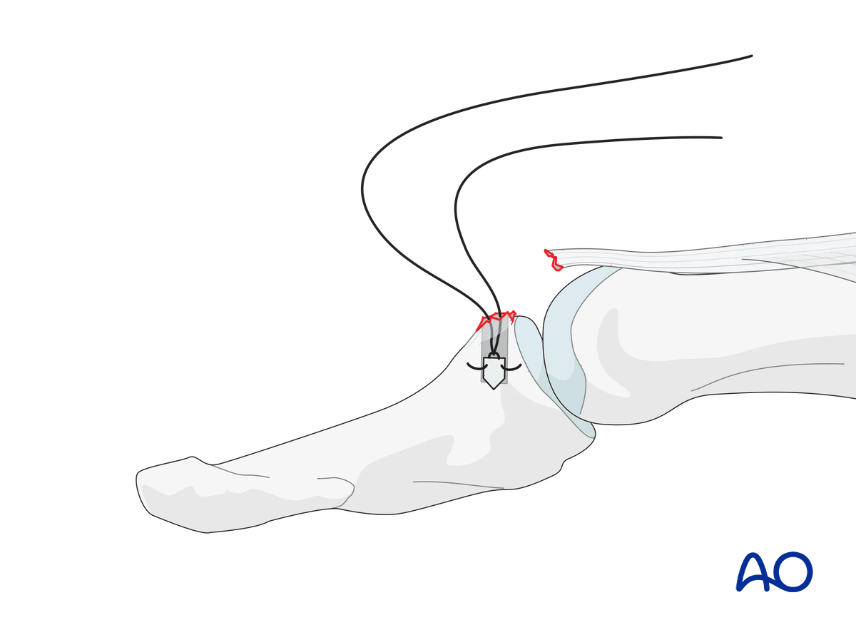
Use 4.0 multifilament nonabsorbable sutures. Tie the sutures around the avulsed fragment.
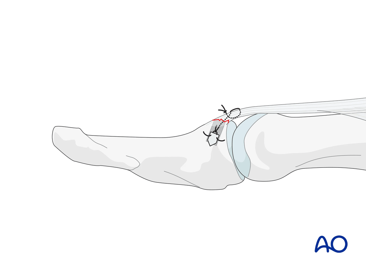
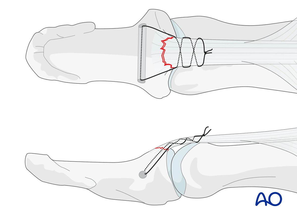
7. Option: transverse intraosseous loop technique
To avoid damage to the nail bed, a transosseous bone tunnel may be used for suture passing.
This tunnel is created with a transdermal needle in the transverse plain of the distal phalangeal base. This is also helpful for passing the suture.
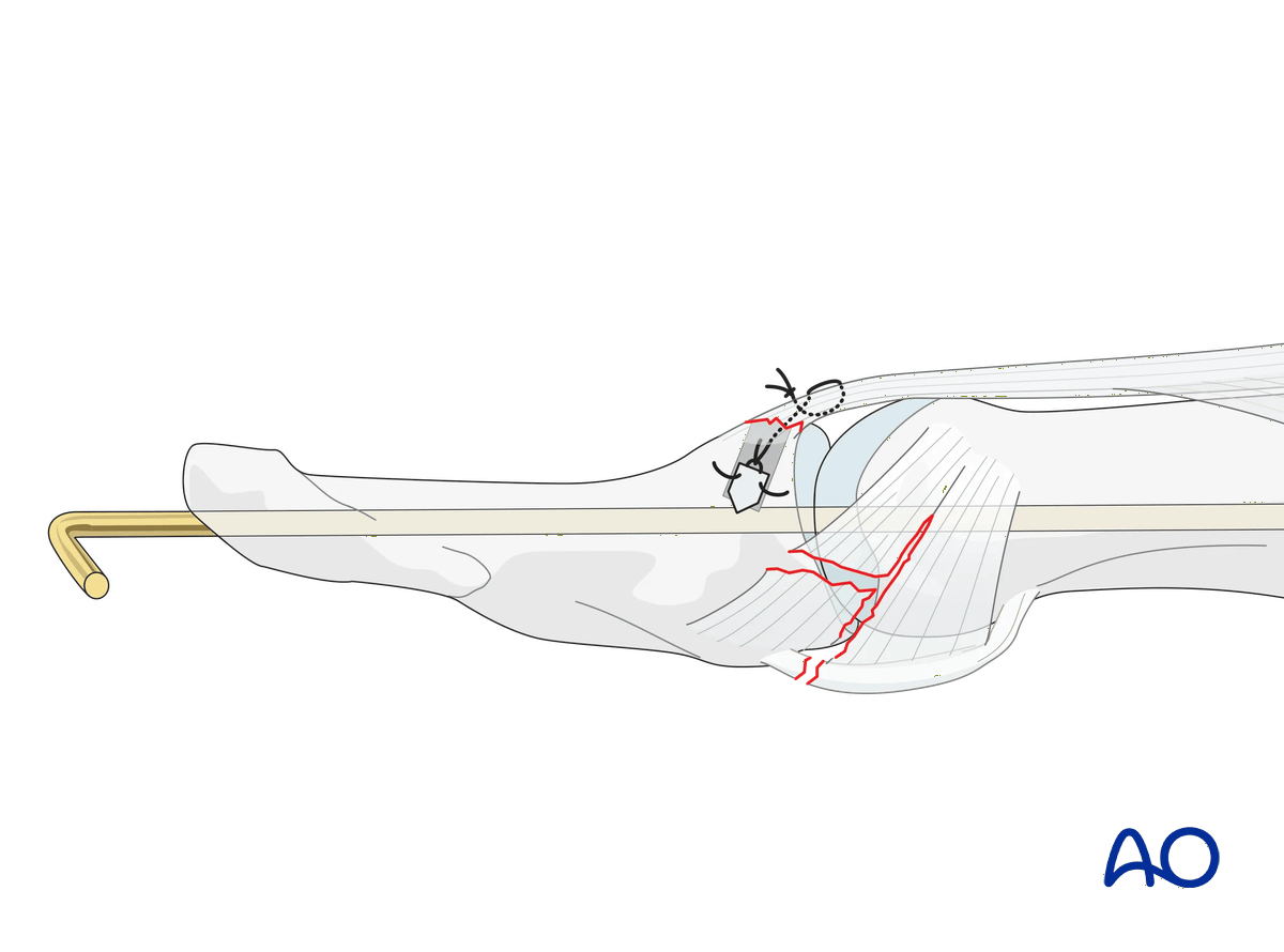
8. Joint transfixation in unstable situations
Palmar subluxation usually persists if the collateral ligament and the volar plate are ruptured.
In such a case, the IP joint needs to be immobilized with a K-wire.
9. Splinting
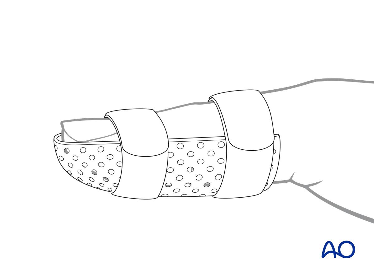
Immobilize the IP joint in extension in a palmar splint or thermoplastic splint leaving the MCP joint free. This also protects a transarticular K-wire from breakage.
A Coban bandage to reduce swelling is recommended.
10. Aftercare
Healing phases
The aftercare can be divided into four phases of healing:
- Inflammatory phase (week 1–3)
- Early repair phase (week 4–6)
- Late repair and early tissue remodeling phase (week 7–12)
- Remodeling and reintegration phase (week 13 onwards)
Full details on each healing phase can be found in the chapter Postoperative treatment of thumb injuries.
Management of swelling
The splint allows for movement at the MCP joint and helps with pain and edema control. The arm should be actively elevated to help reduce the swelling.
Follow-up
A wound check is performed 3–5 days after surgery. Dressings are changed to avoid the tourniquet effect of dried exudate or bleeding within the dressings.
Sutures may be removed after 10 days.
Strict immobilization for 8 weeks is needed for stable healing and good outcome.
After this period, start weaning the splint during daytime. Splinting during nighttime should be continued for an additional 4 weeks.
Mobilization
Hand therapy is recommended to prevent soft-tissue atrophy and joint contracture, which leads to a poor outcome, and subsequent treatment is difficult.
Functional exercises of the nonimmobilized joints should be started immediately to keep uninjured joints mobile.
Active motion of the IP joint is permitted after removal of the K-wire. Passive motion should be postponed for 8–10 weeks to avoid the risk of recurrent mallet deformity.