

Collateral ligament repair
1. General considerations
Introduction
In a dislocation of the interphalangeal (IP) joint, usually, ligaments are ruptured.
Usually, these soft-tissue injuries heal without reconstruction.
Collateral ligament ruptures may need repair if the joint remains unstable after closed reduction.
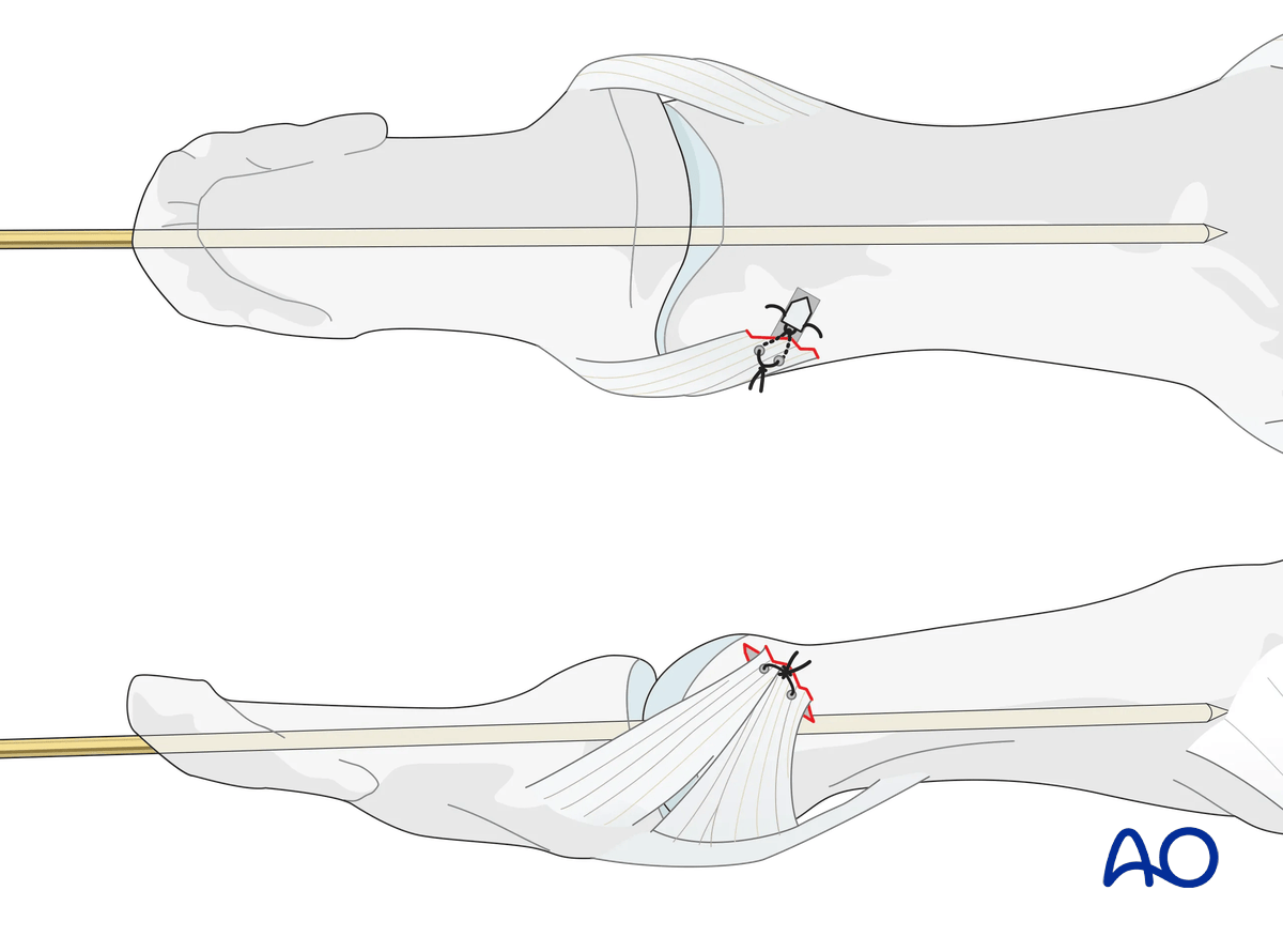
Ligament injuries
The collateral ligament usually tears at one of two locations:
- At its attachment to the proximal phalanx
- At its attachment to the volar plate and distal phalanx
Often, these injuries are accompanied by a partial lesion of the volar plate.
In this procedure, the reattachment to the proximal phalanx is shown.
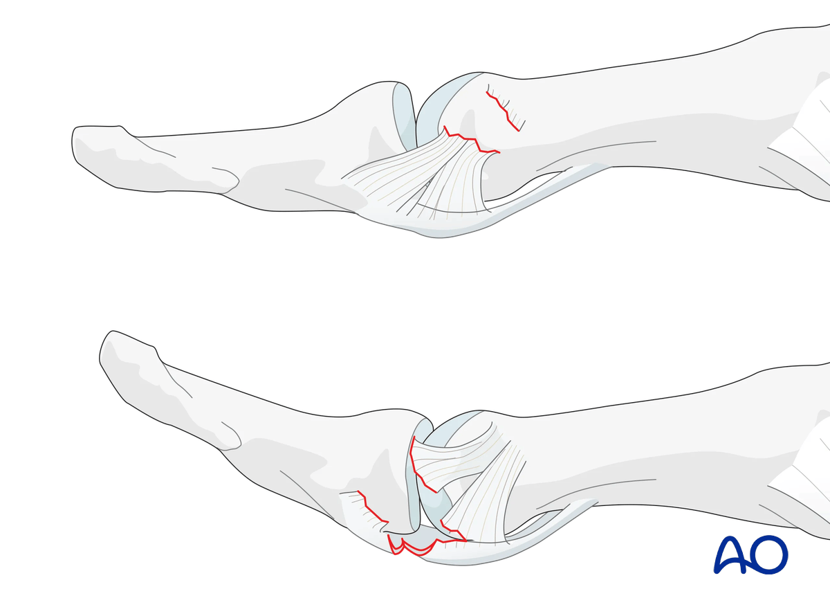
Treatment principles
When ligament repair is necessary, the surgeon should be aware of three guiding principles:
- Know precisely where the lesion is
- Be familiar with the approach and the local anatomy
- Minimizing any soft-tissue dissection
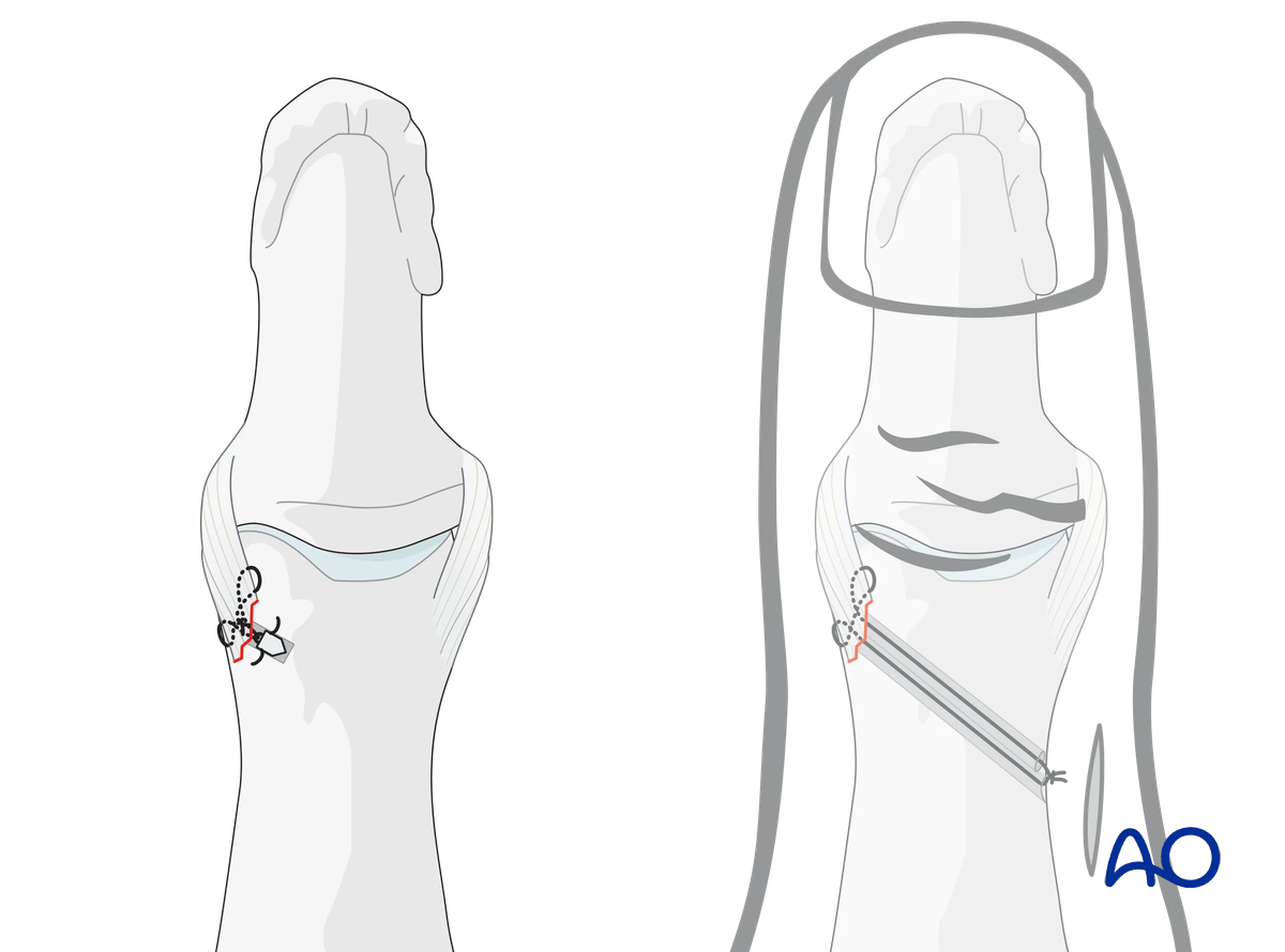
Suture anchors or bone tunneling for repair of ligament detachments
Two alternative techniques are available for collateral ligament reattachment: suture anchors or bone tunneling.
The advantage of suture anchors is the relative ease of the procedure. It is also a time-saving technique.
Tunneling is the more demanding procedure, but it is significantly less expensive.
The details on suture anchoring are shown below.
2. Patient preparation
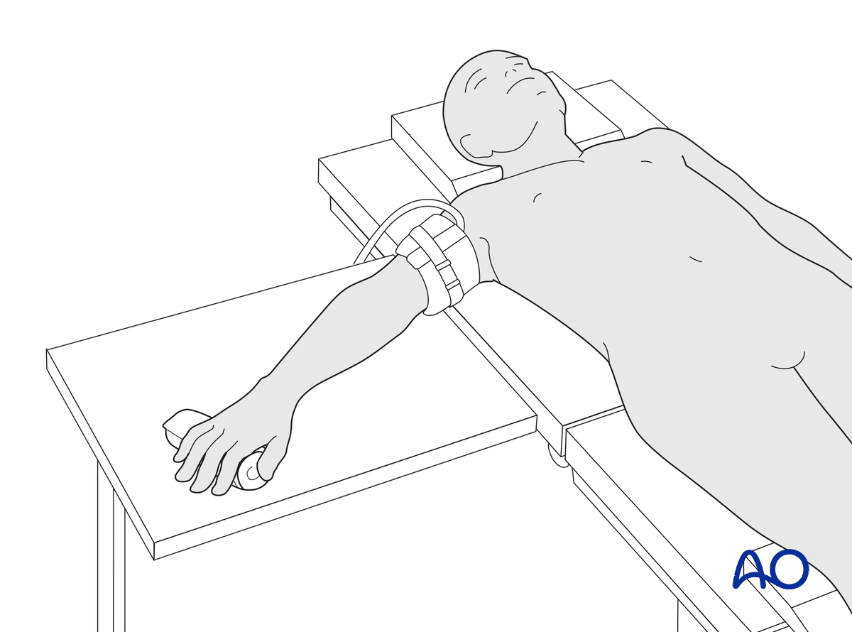
This procedure is usually performed with the patient in a supine position with the arm on a radiolucent side table.
3. Approach
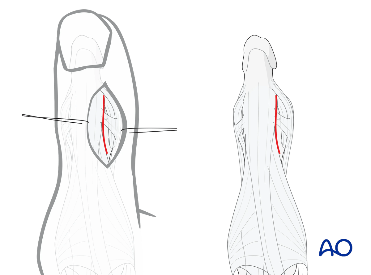
To expose the lateral aspect of the IP joint, perform a straight dorsoradial, or dorsoulnar, skin incision and retract the EPL tendon as described in the dorsal approach to the IP joint.
4. Closed reduction
Apply traction to the thumb, with the IP joint in slight flexion to relax the flexor tendon.
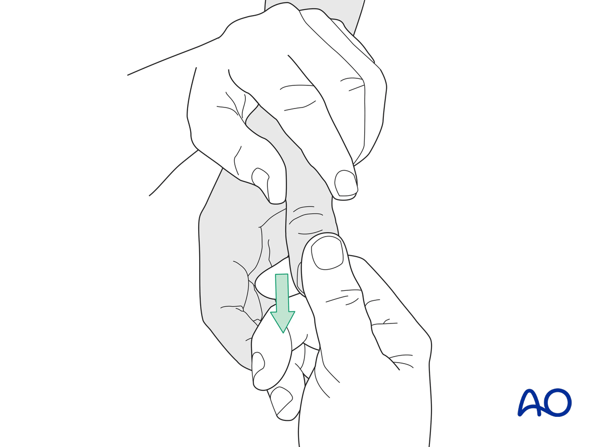
Check reduction with image intensification in two planes. The images should show complete joint congruency.
Check the stability of the joint putting stress on both collateral ligaments.
Check for normal flexor and extensor tendon function at the IP joint.
5. Repair with a suture anchor
Preparation of the attachment site
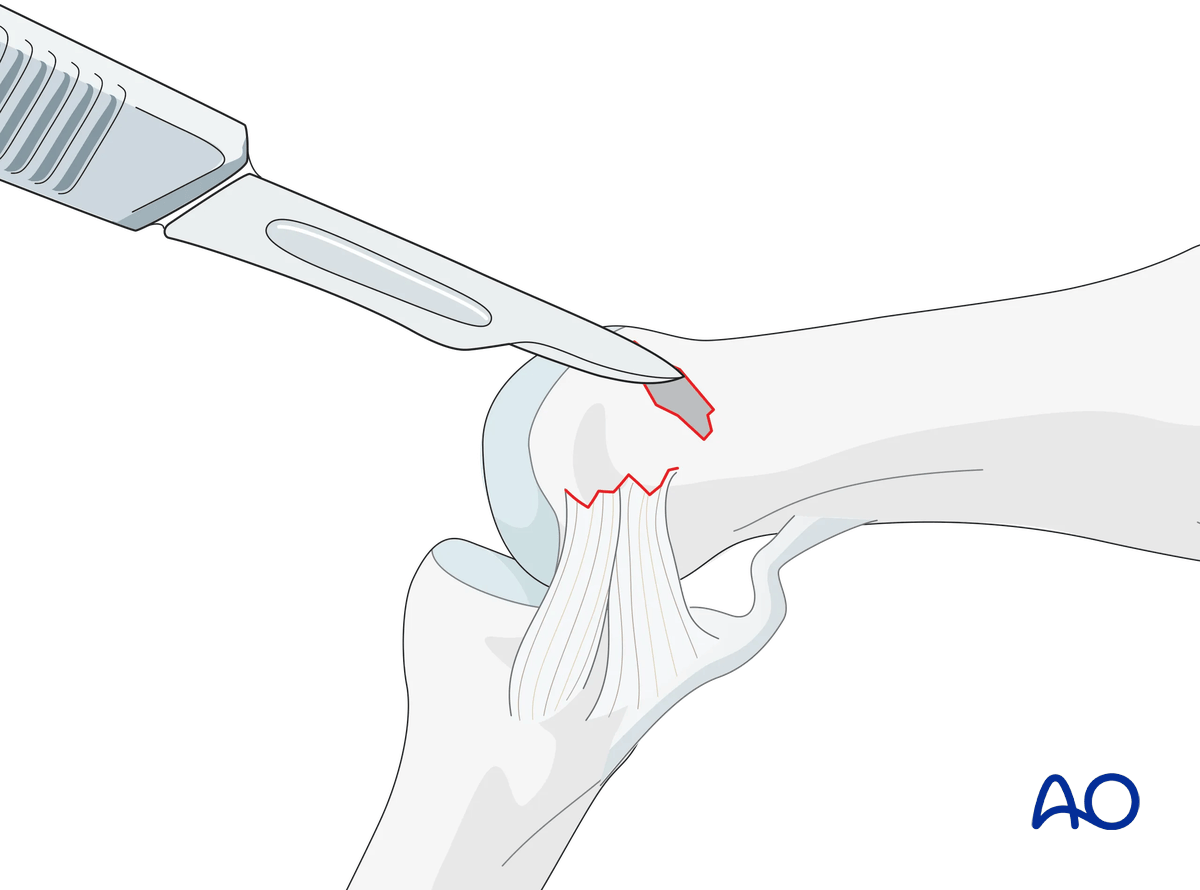
Clean the attachment site of the collateral ligament of any remaining soft tissues with a scalpel, tiny curette, or small burr.
Prepare the reattachment site by exposing the cancellous bone. This improves the vascularity of the site and aids later healing.
Drilling an anchor hole
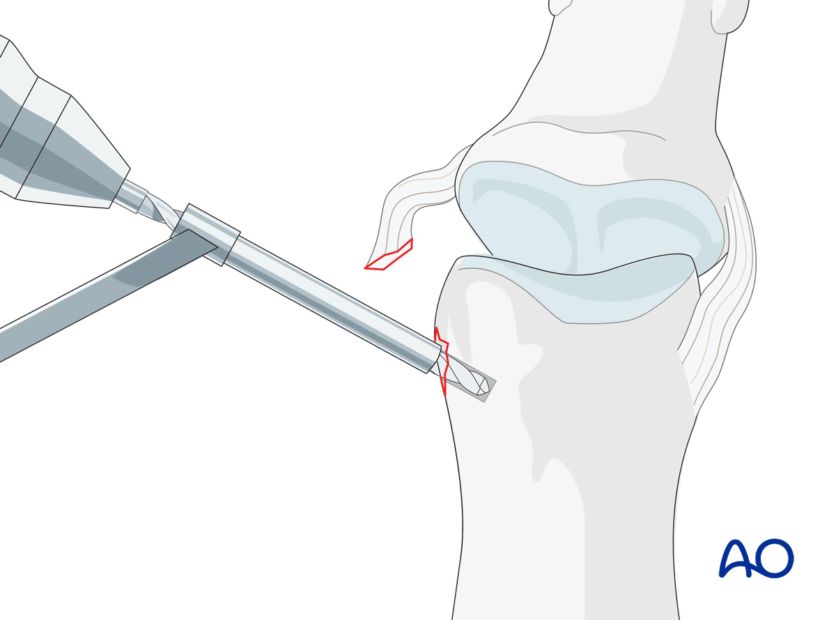
Keep the finger flexed for better exposure.
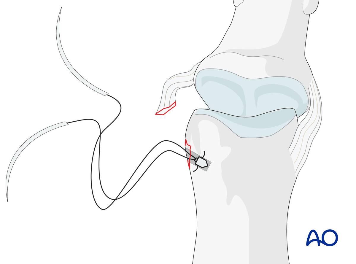
Insert the anchor according to the manufacturer’s instructions at the isometric point of insertion.
Insertion of the anchor
Insert the anchor.
Ensure that the whole anchor is completely buried in the bone.
Insertion of sutures
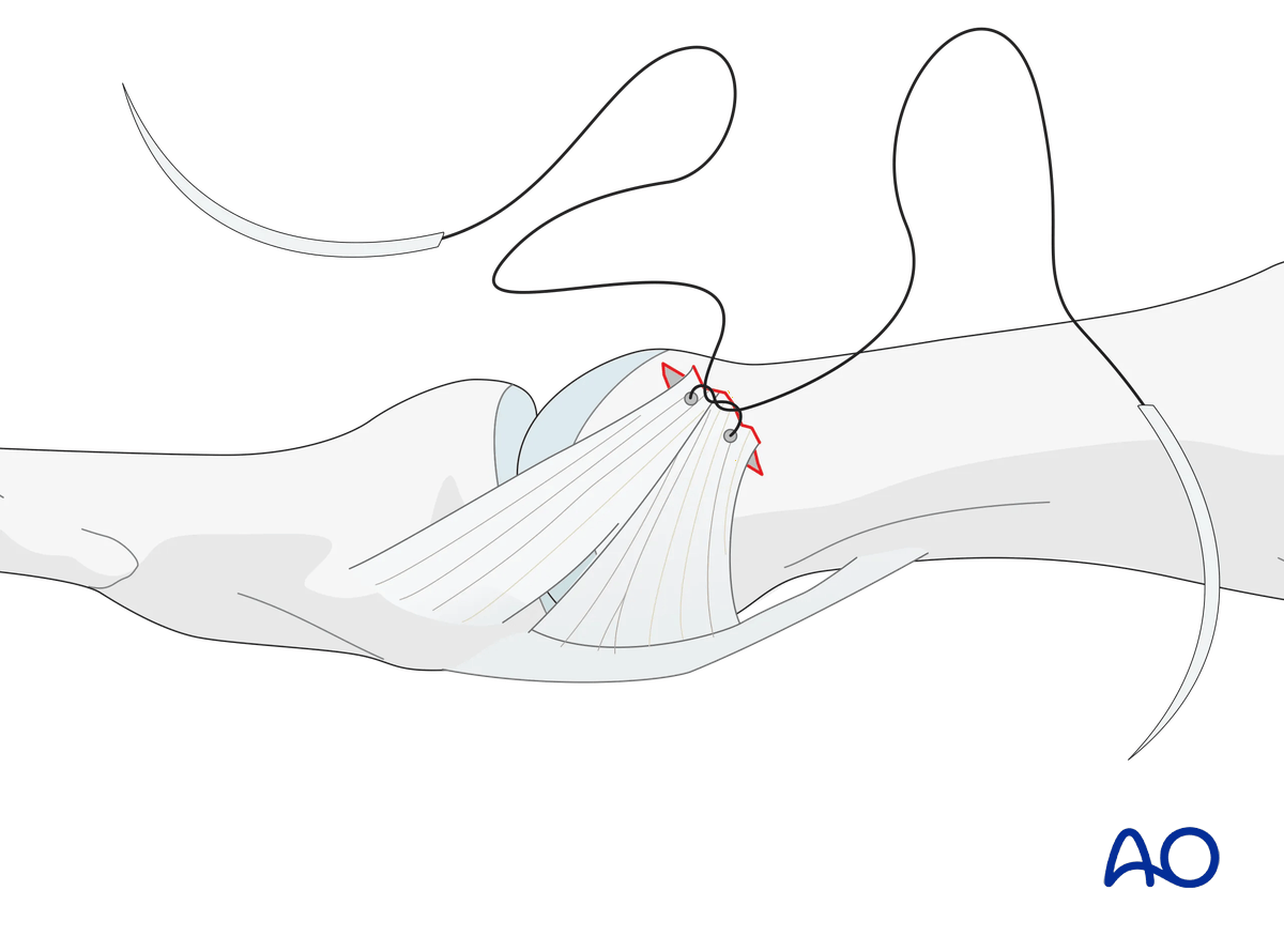
Insert the sutures through the ligament.
Reapproximate the ligament to the phalanx and make a loop at each end of the thread as an anchoring pass. Tie a knot to secure the ligament to the phalanx with the IP joint in extension or slight flexion.
Reattaching the ligament close to the subchondral bone will ensure a smooth surface for ideal mobility.
6. Joint transfixation with K-wire
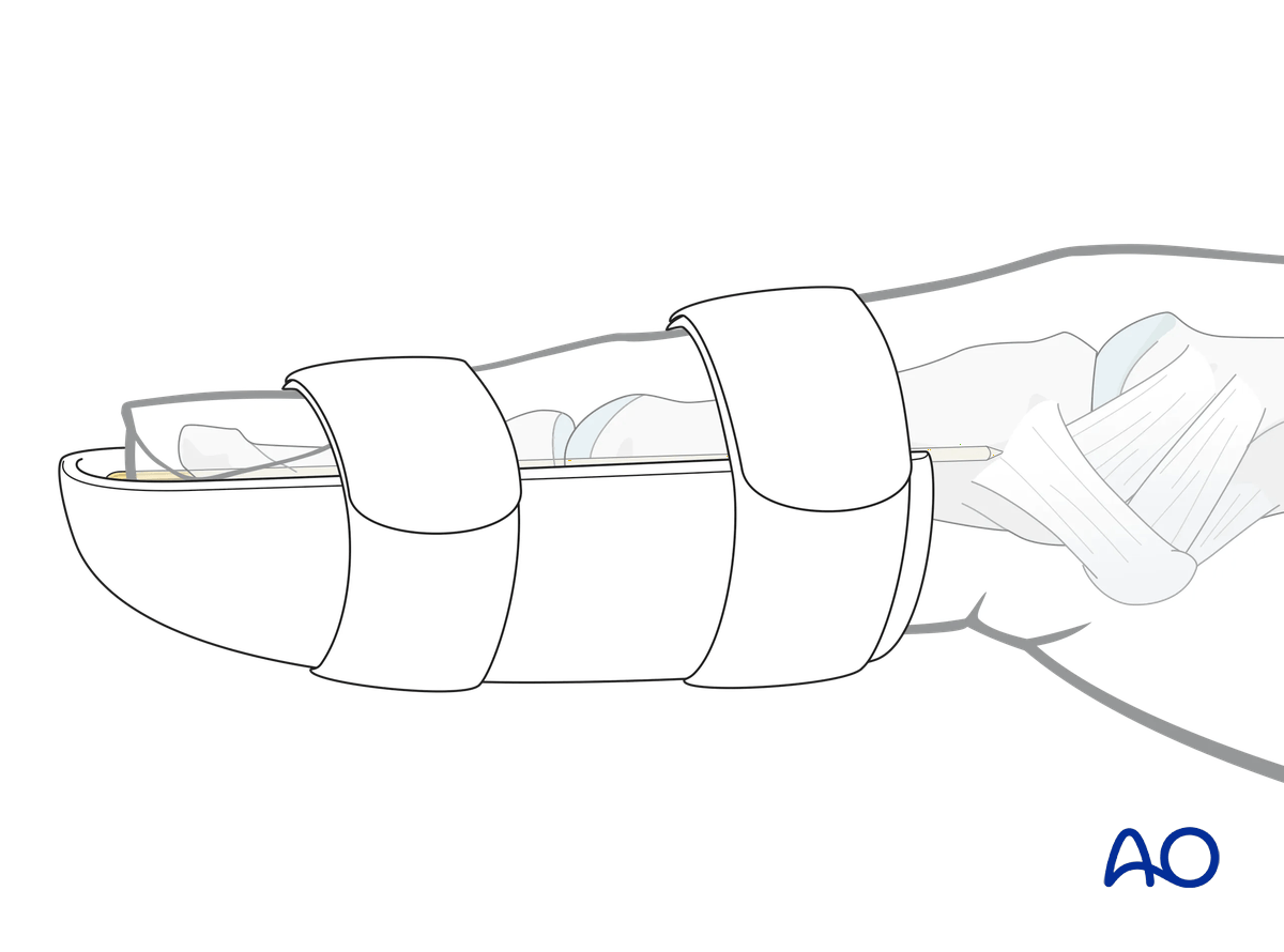
Insert an intramedullary retrograde K-wire across the IP joint to protect the ligament reattachment. Be careful not to penetrate the MCP joint.
Leave the end of the K-wire outside of the skin for later removal.
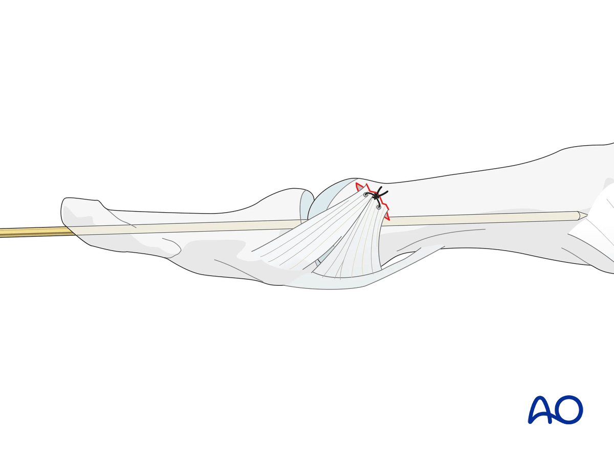
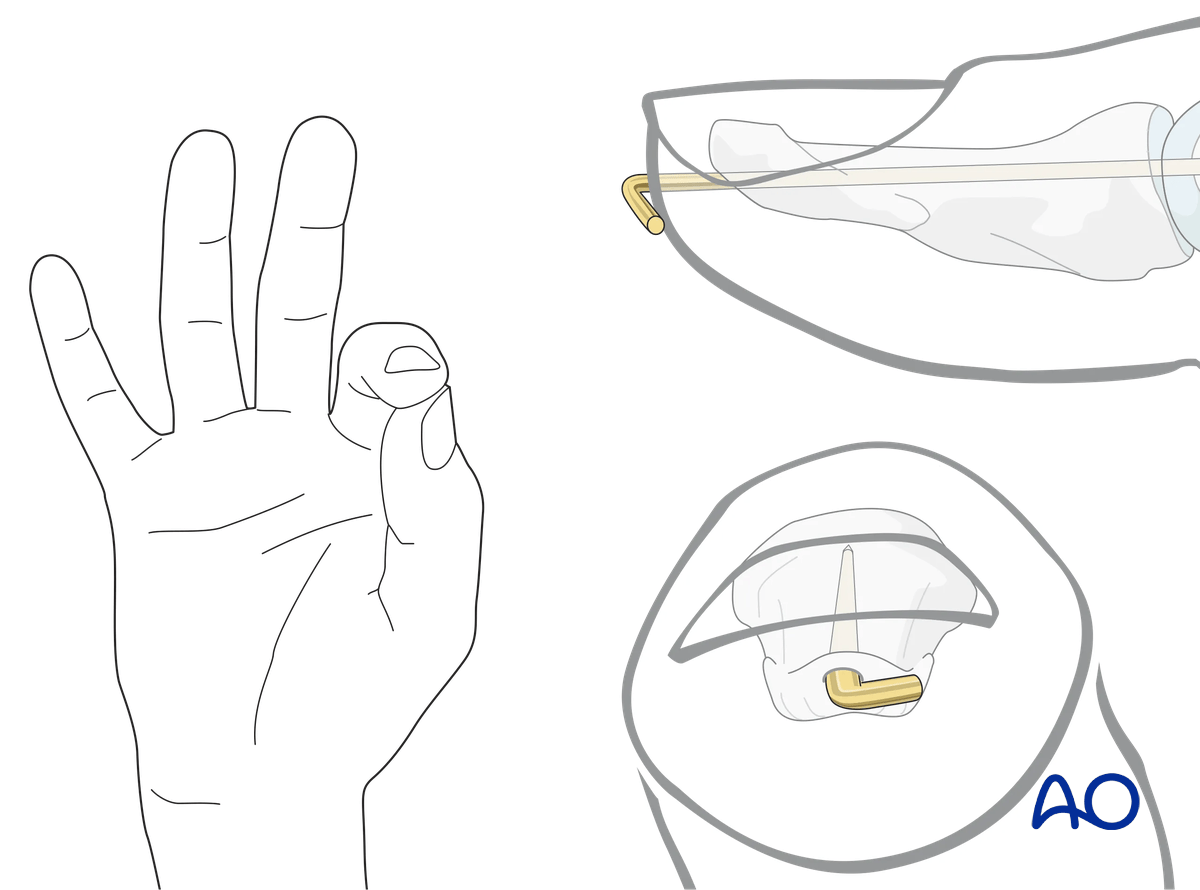
Cut the K-wire so that it protrudes through the skin. Bend its end to form an L-configuration to prevent catching on clothing, etc. The tip of the bent wire can be rotated towards the radial side to avoid interfering with pinch grip, which involves the ulnar side of the pulp.
Leaving the K-wire to protrude through the skin in this way has the advantage of its being easy to remove. The disadvantages are patient discomfort and risk of pin-track infection.
Make sure that the bent end does not irritate or compress the skin.
7. Final assessment
Confirm anatomical reduction and fixation with an image intensifier.
8. Splinting
Apply an IP joint splint, leaving the MCP joint free, for 6 weeks to protect the transarticular K-wire from breakage.
9. Aftercare
Healing phases
The aftercare can be divided into four phases of healing:
- Inflammatory phase (week 1–3)
- Early repair phase (week 4–6)
- Late repair and early tissue remodeling phase (week 7–12)
- Remodeling and reintegration phase (week 13 onwards)
Full details on each healing phase can be found in the chapter Postoperative treatment of thumb injuries.
Management of swelling
The splint allows for movement at the MCP joint and helps with pain and edema control. The arm should be actively elevated to help reduce the swelling.
Mobilization
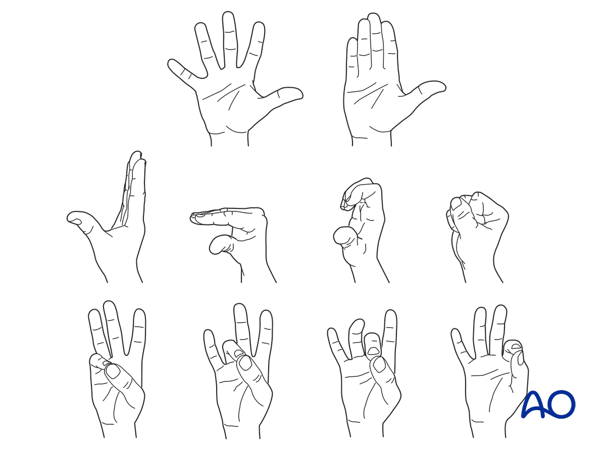
To prevent joint stiffness, the patient should be instructed to begin active motion (flexion and extension) of all nonimmobilized joints of the hand immediately after surgery.
Follow-up
See the patient 6 weeks after surgery for K-wire removal.


