

Screw fixation
1. General considerations
Introduction
A dorsal avulsion of the distal phalangeal base can be fixed with a screw if the proximal fragment is large enough and comprises more than half of the articular surface.
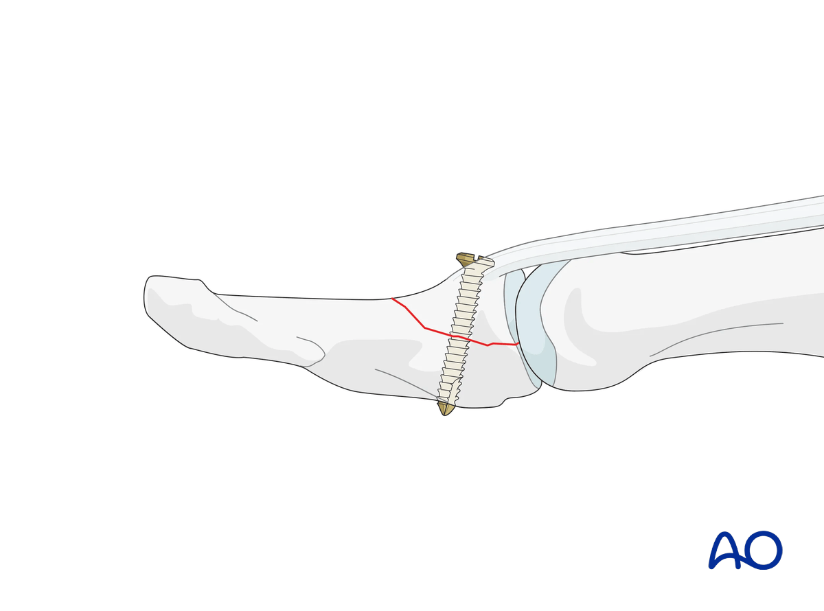
Lag screw vs position screw
Principally, the fracture should be fixed with a lag screw technique. In the metaphyseal parts of the phalanges, it is usually not advisable to drill a gliding hole through the small metaphyseal bone. A position screw holding a compression achieved with reduction forceps provides sufficient stability.
Operative treatment: caveat
Indications for surgical intervention are:
- Open fractures
- Palmar subluxation of the IP joint

Surgical management of these fractures is difficult and has many potential complications. The soft tissues are only precariously vascularized, and the fragments are very small and prone to further comminution. Healing can often be slow.
2. Surgical anatomy
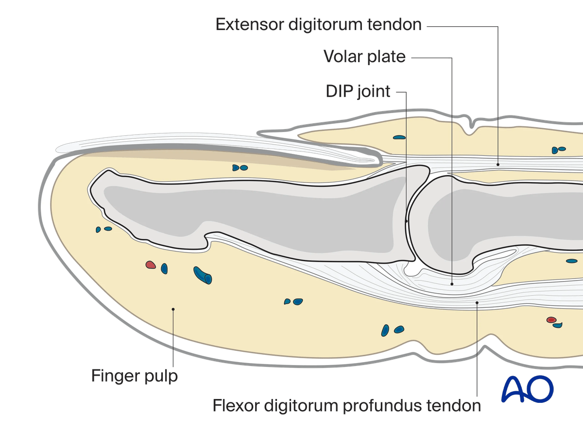
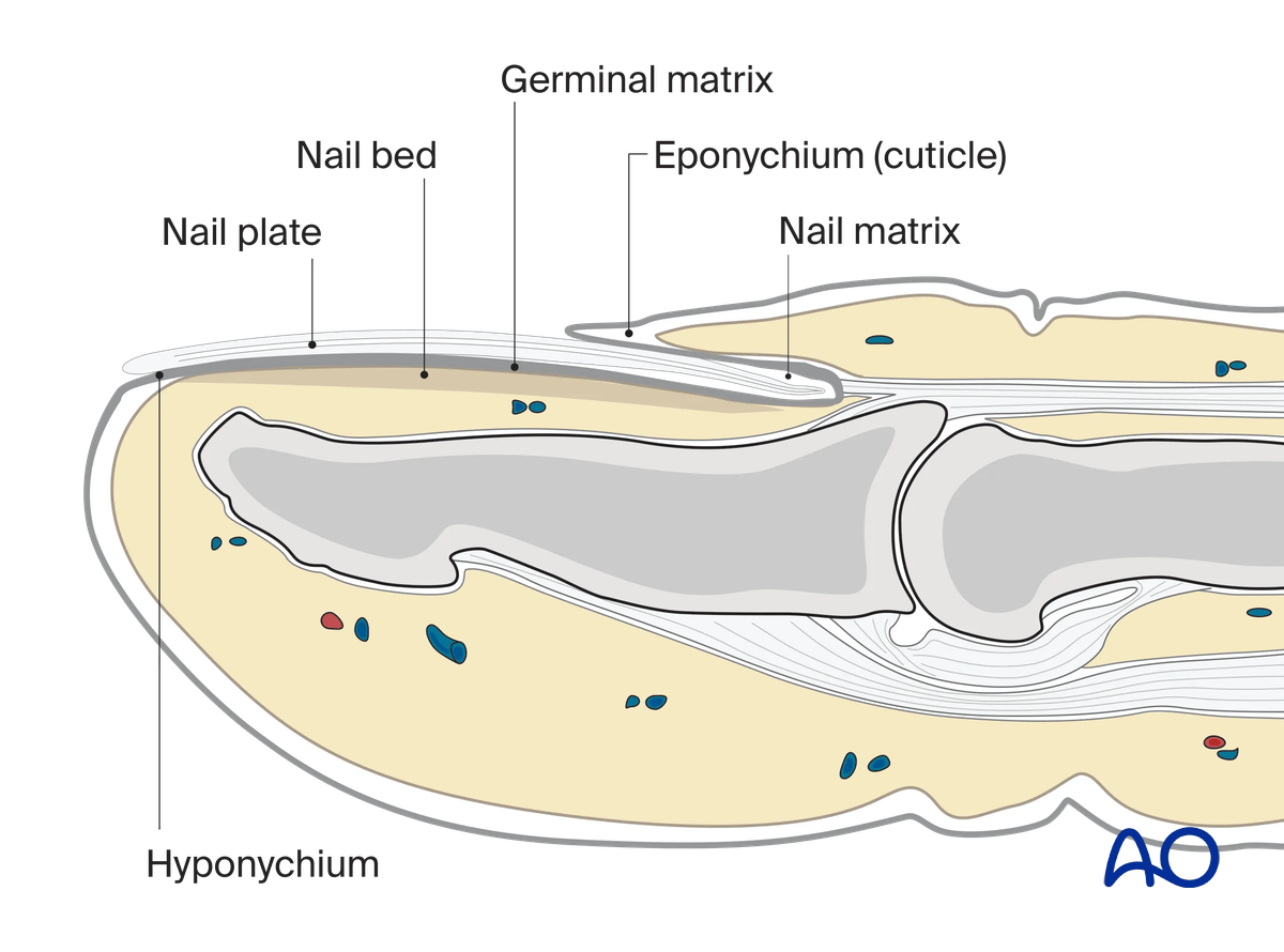
The base of the distal phalanx has a prominent dorsal crest at the insertion of the extensor tendon. The tendon is also adherent to the interphalangeal (IP) joint capsule.
On the palmar surface is the insertion of the flexor pollicis longus tendon.
The flexor tendon occupies the whole width of the base of the distal phalanx. It is made up of two different fibers. The superficial fibers attach to the lateral aspects of the phalanx. The deep fibers run centrally and attach more distal in the palmar aspect of the phalanx.
The volar plate is very flexible, allowing hyperextension of the IP joint and pulp-to-pulp pinch.
The vascularity of the extensor tendons is more precarious than that of the flexor tendons. This prolongs extensor tendon healing time.
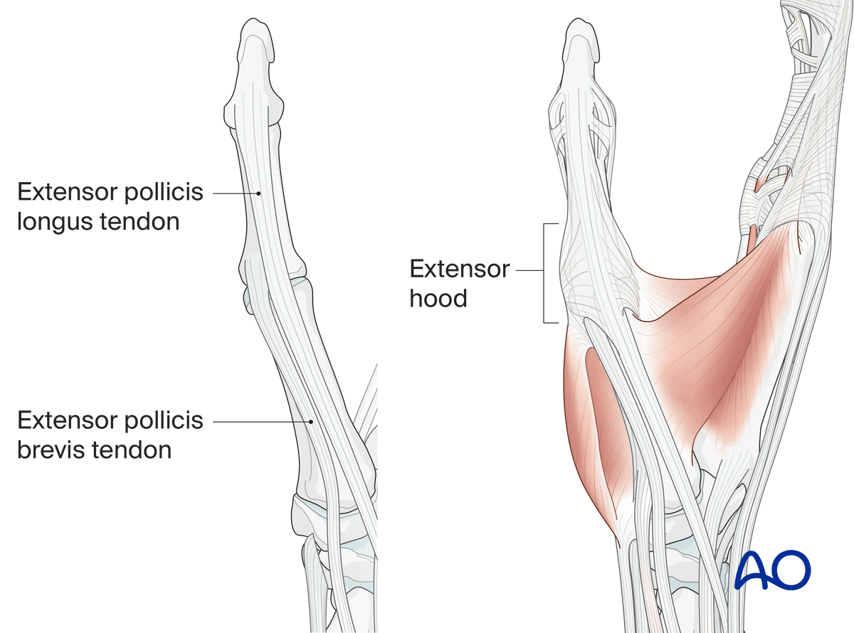
The extensor hood of the thumb consists of the radially located extensor pollicis brevis tendon (insertion: base of the proximal phalanx) and the ulnarly located extensor pollicis longus tendon (insertion at the base of the distal phalanx).
Both tendons are held in position by the sagittal bands at the level of the MCP joint and the extensor hood at the level of the proximal phalanx.
At the level of the MCP joint the abductor pollicis aponeurosis radiates into the extensor hood.
3. Patient preparation
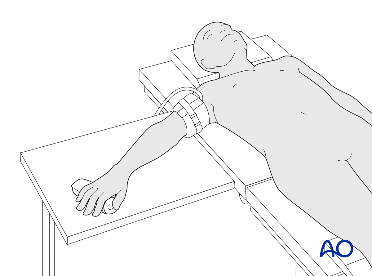
This procedure is usually performed with the patient in a supine position with the arm on a radiolucent side table.
4. Approach
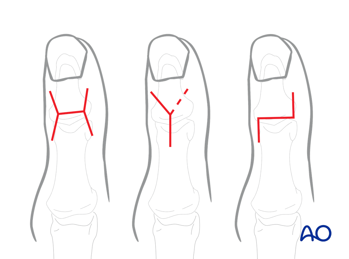
For this procedure, a dorsal approach to the IP joint is typically used.
5. Reduction
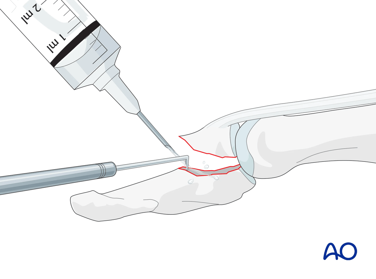
Flex the IP joint. To better visualize the fracture, use a syringe to clean out blood clots with a jet of water.
Often the degree of comminution is not apparent from the x-rays and can only be determined under direct vision.
Use a dental pick to carefully free interposed tissues and remove blood clots and other debris.
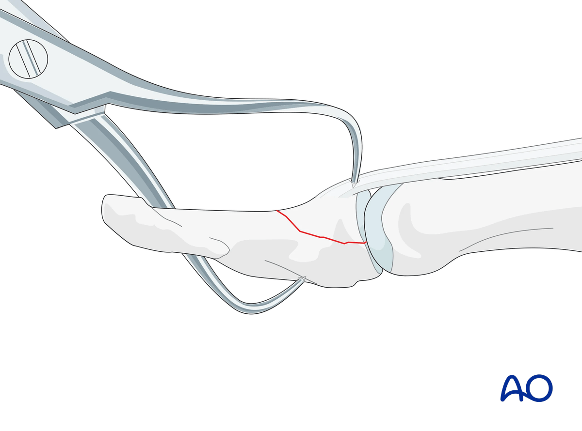
Extend the IP joint. Reduce the fragment with percutaneous reduction forceps.
6. Fixation
Selecting screw size
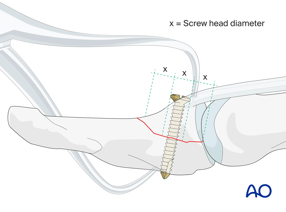
The maximal permitted diameter of the screw head is one-third of the diameter of the avulsed fragment.
The screw length must be adequate for the screw to engage the opposite cortex.
Most commonly, a 1.0 mm diameter screw is used. 1.3 mm screws can also be used for large fragments.
Drilling
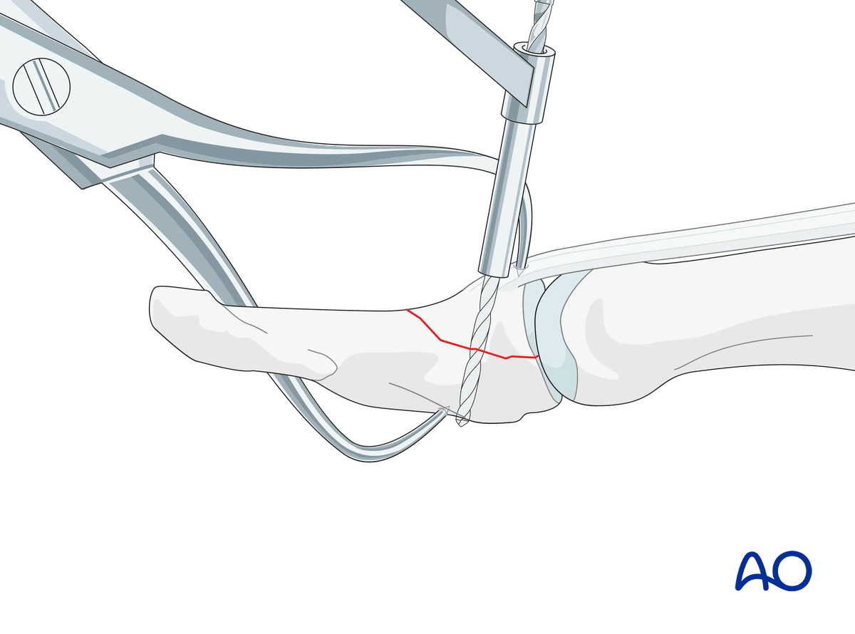
Drill a hole in both cortices.
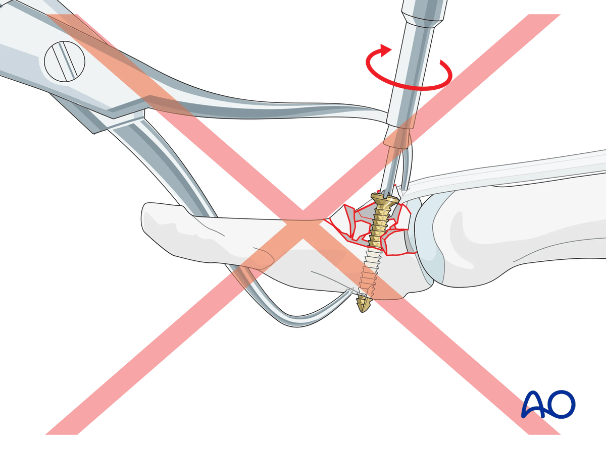
Countersink the near cortex slightly to allow the screw head to sink almost flush with the cortical surface. This facilitates radial preload of the cortex to minimize the risk of secondary fracture and loosening of the screw. Be careful not to over-countersink the near cortex.
Determining the screw length
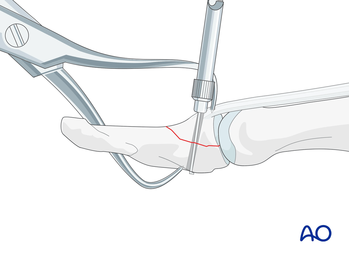
Use a depth gauge to determine the screw length.
Screw insertion
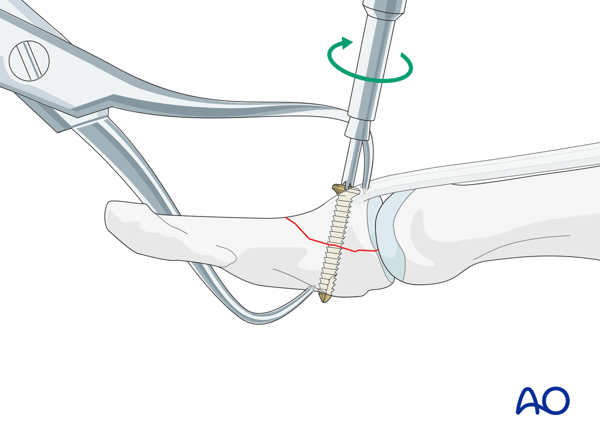
Insert a self-tapping screw and tighten it. The screw should engage the opposite cortex. Be careful not to overtighten the screw to avoid stripping the threads with subsequent loss of fixation.
Final check
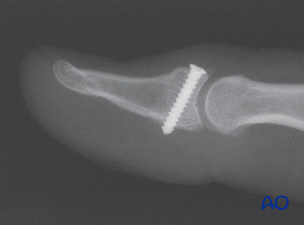
Confirm articular reduction and correct screw placement with an image intensifier. Reduction must be anatomical.
Option in large fragments: 2nd screw
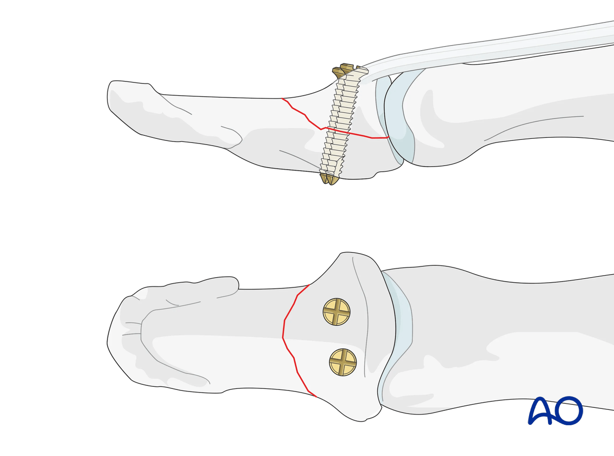
A second screw can be added in very large fragments to improve stability.
7. Joint transfixation in unstable situations
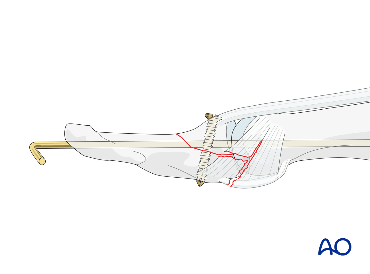
Palmar subluxation usually persists if the collateral ligament and the volar plate are ruptured.
In such a case, the IP joint needs to be immobilized with a K-wire with a splint protecting the wire from breakage.
8. Splinting
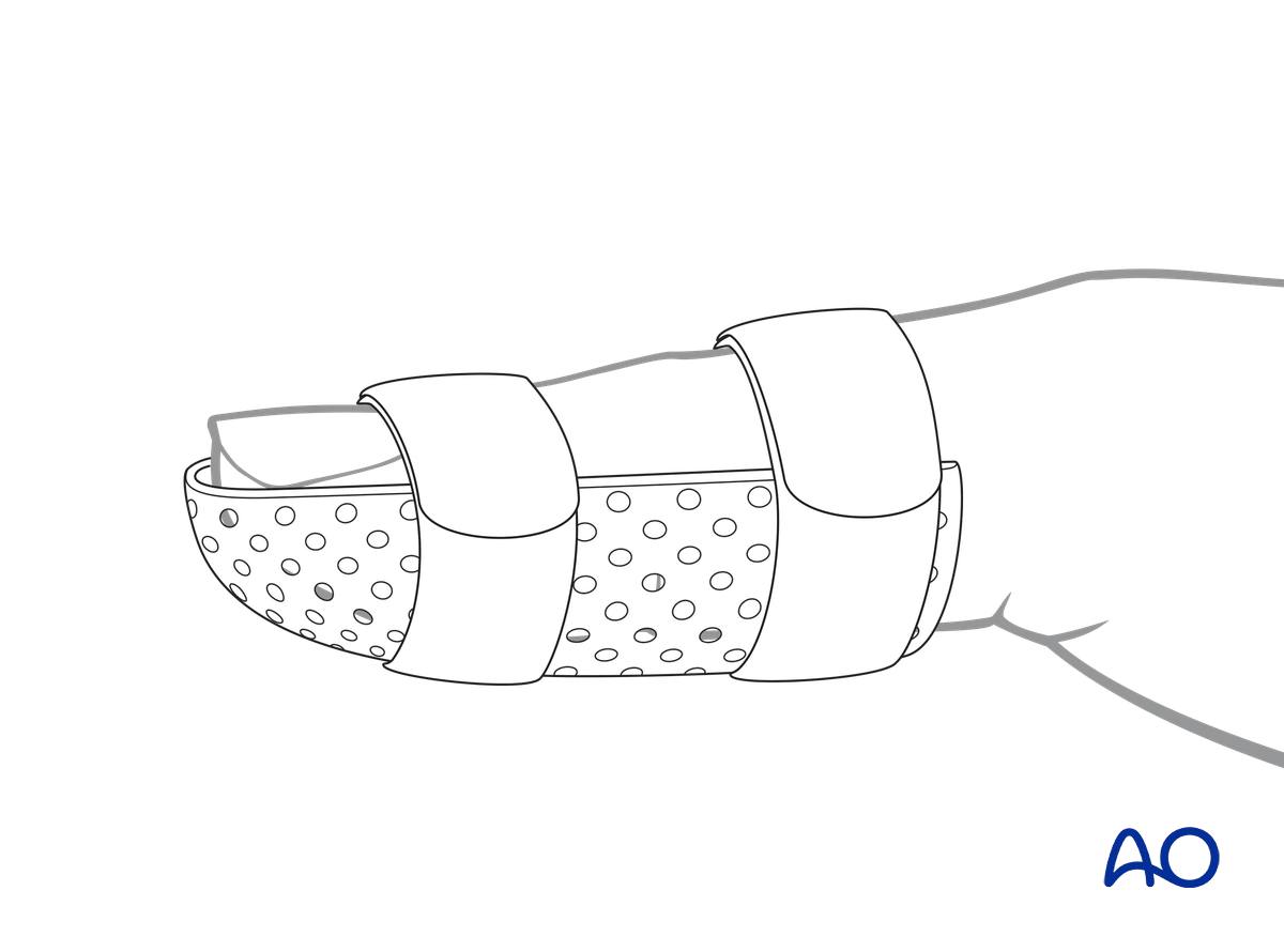
Immobilize the IP joint in extension in a palmar splint or thermoplastic splint leaving the MCP joint free. this also protects a K-wire.
A Coban bandage to reduce swelling is recommended.
9. Aftercare
Healing phases
The aftercare can be divided into four phases of healing:
- Inflammatory phase (week 1–3)
- Early repair phase (week 4–6)
- Late repair and early tissue remodeling phase (week 7–12)
- Remodeling and reintegration phase (week 13 onwards)
Full details on each healing phase can be found in the chapter Postoperative treatment of thumb injuries.
Postoperative management
A wound check is performed 3–5 days after surgery. Dressings are changed to avoid the tourniquet effect of dried exudate or bleeding within the dressings.
Sutures may be removed after 10 days.
Management of swelling
The splint allows for movement at the MCP joint and helps with pain and edema control. The arm should be actively elevated to help reduce the swelling.
Mobilization
Mobilization depends on the stability gained by the fixation.
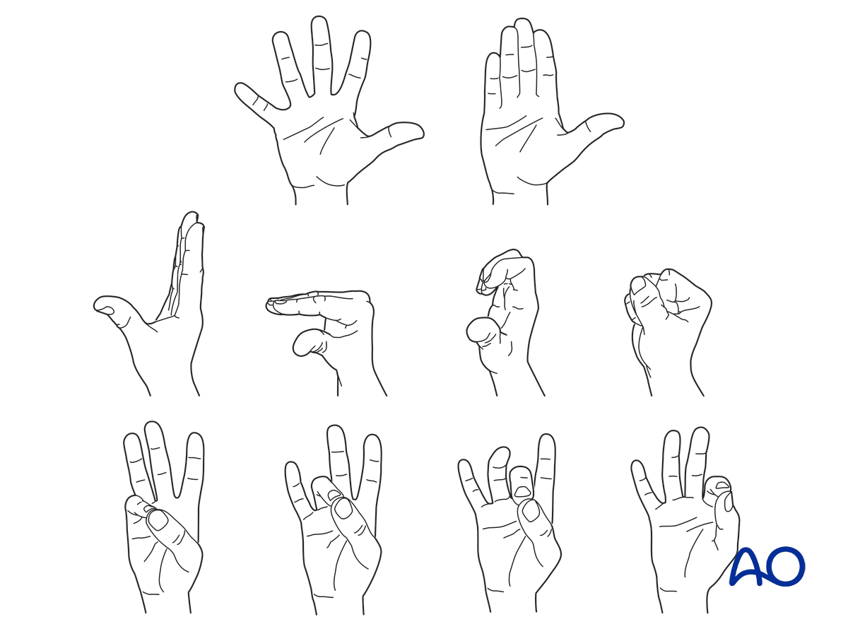
Hand therapy is recommended to prevent soft-tissue atrophy and joint contracture, which leads to a poor outcome, and subsequent treatment is difficult.
If the fixation is strong enough and the IP joint is not transfixed, the patient is encouraged to take off the splint 2–3 times daily and commence with gentle active exercises.
Follow-up
After 6 weeks, an x-ray is taken to confirm healing. Splinting can then usually be discontinued.
If, after 6 weeks, x-rays confirm healing and the patient is painless, full loading can be permitted.


