MIO - Lag screw fixation
1. Principles/Introduction
Main goal
In this procedure the main goal is to reconstruct the joint surface.
Screw selection
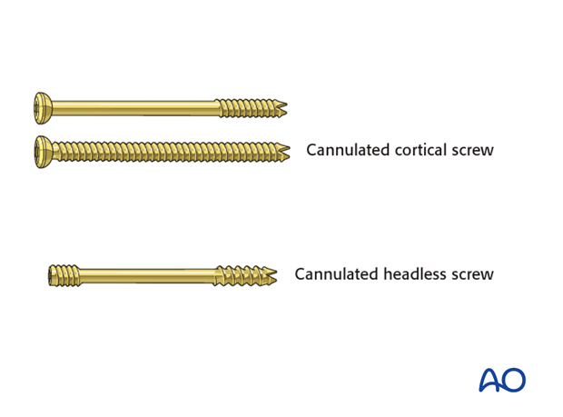
For this procedure the following screws are typically used:
- Cannulated 3.5 lag screws
- Headless 3.0 screws
Headless screws are preferable where fixation involves inserting them through capsule or labrum.
If a headless screw is used, bicortical purchase is preferred but not required for compression.
2. Patient preparation
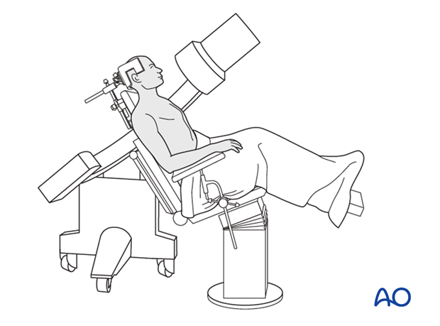
Depending on the approach, this procedure may be performed with the patient in a beach chair position or lateral position.
3. Approach
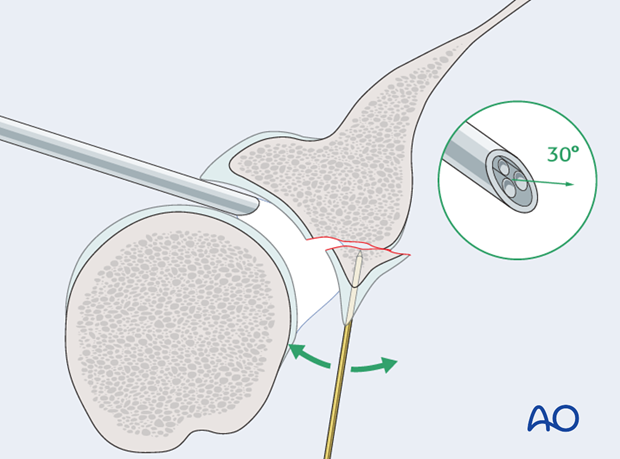
Anterior fragments are reduced and fixed through the minimal invasive anterior approach with the camera inserted posteriorly.
4. Reduction
Reduction of the articular surface may be facilitated by the insertion of a K-wire to be used as joystick. For this reason, we prefer to use the cannulated system and insert the K-wire in such a way that it will subsequently serve as a guide for the lag screw trajectory.
The liquid used for distension should not be under high pressure. If it is under high pressure it will drive the fragments apart and make the reduction difficult.
If a satisfactory reduction cannot be achieved by closed manipulation one must resort to an open reduction and fixation.
Whenever K-wires are used as joysticks, whether as subsequent guide wires or not, they should not be inserted trans articular.

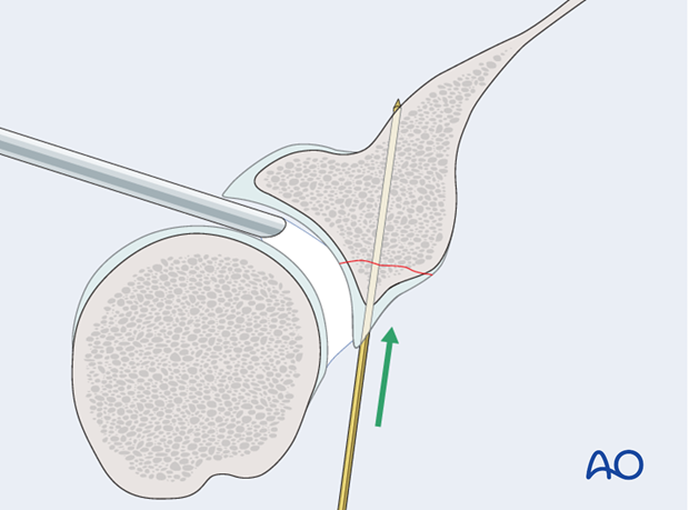
When reduction is completed, the joystick K-wire is now inserted to temporarily fix the fracture.
The pressure of the arthroscopy liquid can now be increased to normal if necessary.
Make sure that K-wires are not directed into the suprascapular notch where they can compromise the neurovascular bundle.
5. Fixation
An appropriate length lag screw is inserted.
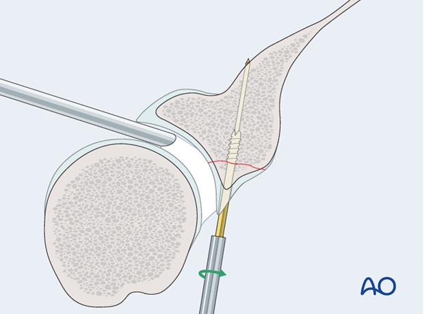
The K-wire is removed.
Depending on the size of the fragment, two lag screws are preferred since one does not offer rotational stability.
Correct reduction is continuously verified by the arthroscopic view.
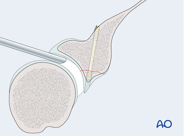
Check the position of the screws and the reduction by image intensification.
6. Aftercare
The aftercare can be divided into 4 phases:
- Inflammatory phase (week 1–3)
- Early repair phase (week 4–6)
- Late repair and early tissue remodeling phase (week 7–12)
- Remodeling and reintegration phase (week 13 onwards)
Full details on each phase can be found here.


