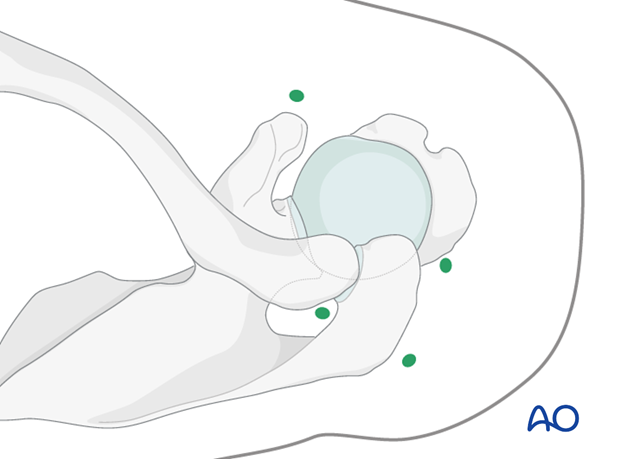
Arthroscopic entry points in the scapula
1. Introduction
An arthroscope is of use only if one is dealing with a fracture that involves the articular surfaces and can be used only if the patient is in the beach chair position.
2. Portals
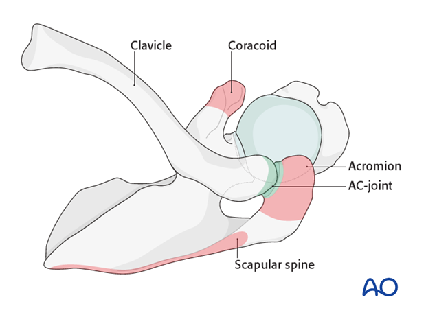
The following bony landmarks are identified by palpation and marked on the patient with a sterile pen.
- Acromion
- Scapular spine
- Coracoid
- AC joint
- Clavicle
The camera is inserted on the opposite side of the main fracture fragment through one of the three standard portals.
3. Dorsal standard portal
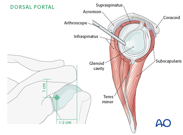
The dorsal standard portal is at the soft spot 1-2 cm medial to the lateral edge of the acromion and 1 cm below the scapular spine.
This portal is used for anterior fractures of the glenoid.
4. Anterior standard portal
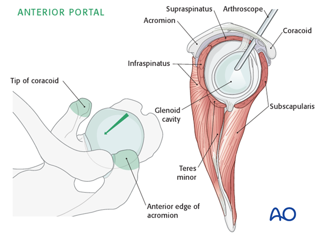
The anterior portal is located between the tip of the coracoid and the anterior edge of the acromion.
This portal is used for posterior fractures of the glenoid.
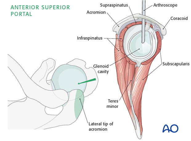
5. Anterior superior standard portal
The anterior superior portal is located just inferior to the lateral tip of the acromion.
This portal is used for inferior fractures of the glenoid.
6. Preparation of camera portal
At the designated spot depending on which portal you are developing make a 1 cm skin incision. Then using blunt dissection with scissors, carry the exposure to the joint capsule.
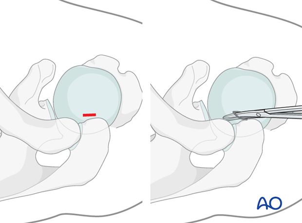
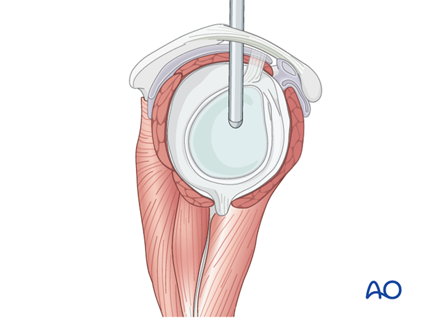
Once you have reached the capsule, take the blunt rod and push it into the joint. Once you are inside the joint enlarge the exposure by inserting a trochar into the joint following closely the rod.
7. Preparation of second portal
The location of the second portal is dictated by the fracture location and from where hardware needs to be inserted. This is the instrument portal.
In the case of an anterior fracture, insert a needle through the described location of the anterior portal. Monitor the entry of the needle through the capsule into the joint. The needle entry into the capsule is monitored through the camera which is previously inserted through a portal on the opposite side of the fracture.
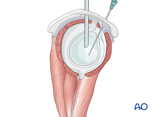
If necessary, additional portals can be used as shown in the illustration.
