ORIF - Plate fixation
1. Introduction
The complex articular fracture is often difficult to reduce. The best provisional reduction, if it cannot be achieved with screws, will now be maintained by the fixation of the plate to the articular block.
Preoperative assessment of these fractures is critical. If the CT and 3D reconstruction suggests that a satisfactory articular reduction of the articular fracture cannot be achieved, then closed treatment may be the best option.
In a patient for whom restoration of function is important, at least 70% of the glenoid should be reconstructable to achieve stability of the shoulder and congruency of the joint.
If the articular segment is reducible, then carry on as for simple articular neck multifragmentary fractures.
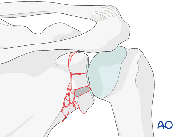
To illustrate the principles, we will here show the treatment of a fracture with but a multifragmentary articular block and multifragmentary neck.
Principle
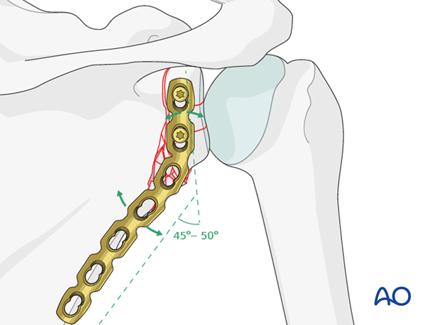
In this procedure the angle subtended between the glenoid and the lateral border of the scapula must be restored.
Hardware selection
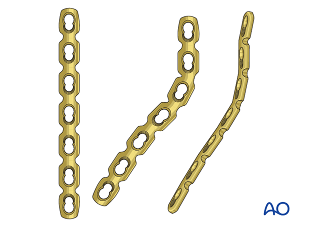
The object is to secure good fixation of the plate to the articular segment, restore the glenoid angle and achieve secure fixation to the lateral border of the scapula. The plate functions as a bridge plate. Its best fixation to the proximal segment is achieved through angular stability and the same applies for its fixation to the lateral border of the scapula.
The best implant is a reconstruction plate which has been appropriately contoured, or a pre-contoured plate. Its fixation to the bone is accomplished with locking screws.
2. Patient preparation
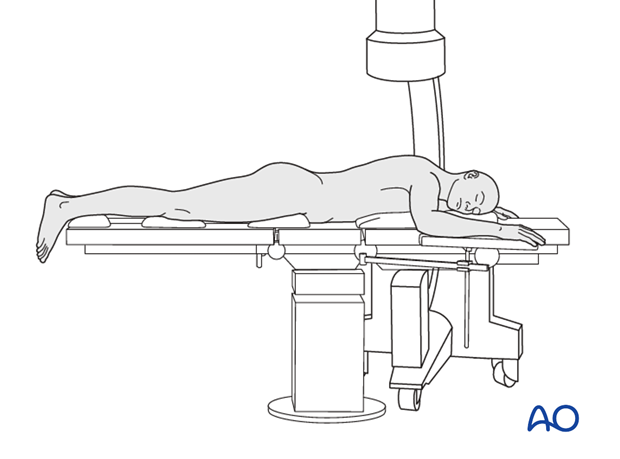
This procedure may be performed with the patient in either a prone position or lateral decubitus position.
3. Approach
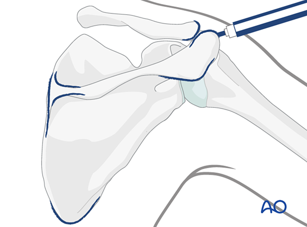
Plating of the glenoid neck and the scapula are performed through a dorsal approach.
4. Reduction and fixation
Reduction
If the fracture is considered reducible, insert a K-wire in a major segment of the glenoid fossa and use it as a joystick for reduction. If possible, it is advantageous to start the reconstruction from either the top, or alternatively a bottom segment.
Note: Careful attention must be made to the orientation of the first main segment as this will determine the angle of the glenoid fossa. Large deviations of the angle (> 20 °) will lead to a reduced function of the shoulder.
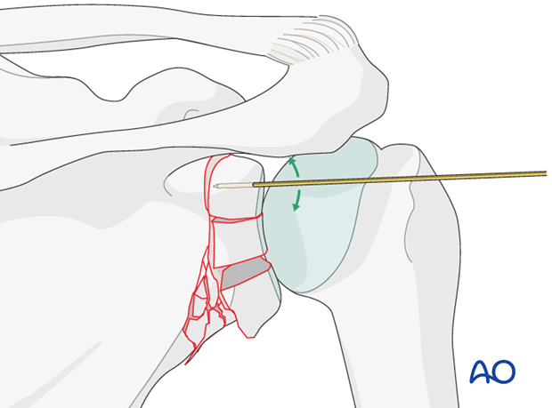
When reduction is achieved, the segment is temporarily fixed to the scapular body by further insertion of the K-wire.
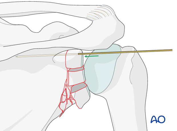
The mid-segment is then reduced.
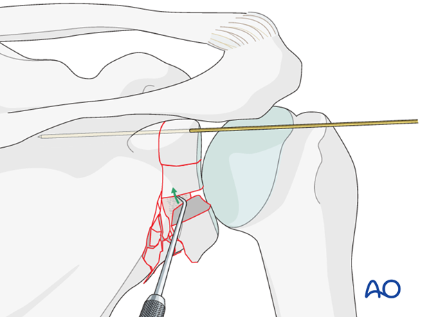
The inferior segment is reduced and temporary fixed with a K-wire. The main goal is to re-establish an anatomical correct articular surface.
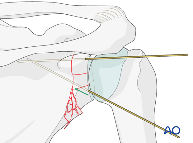
Plate fixation
The plate is bent at the right spot to subtend the usual angle between the glenoid and the lateral border of the scapula (130 - 135 degrees).
When angularly stable fixation is used the plate does not have to fit the bone, but must fit the overall contour.
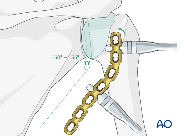
Begin the fixation by attaching the plate to the articular segment.
In the glenoid joint block there is usually space for 2 screws only. These should be placed in the main fragments.
Care must be taken regarding the direction of the locking head screws so that they do not enter the joint.
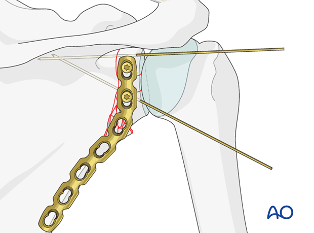
A trans-glenoid X-ray is recorded to verify whether the anatomical angle is achieved.
Large deviations of the angle (>20 °) will reduce the functional outcome.
If need, the K-wires are removed and the plate used as a reduction aid.
When the correct reduction and the correct angle between the glenoid fossa and the lateral border of the scapula have been achieved, screws are inserted along the lateral border of the scapula.
If indicated, use locking screws and in the articular block beware that they do not enter the joint.
Ideally, three bicortical screws are inserted in the lateral scapular border.
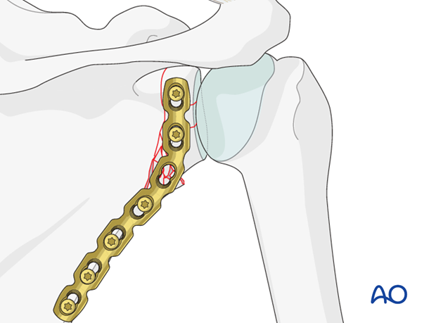
Correct reduction and fixation is verified by image intensification.
5. Aftercare
The aftercare can be divided into 4 phases:
- Inflammatory phase (week 1–3)
- Early repair phase (week 4–6)
- Late repair and early tissue remodeling phase (week 7–12)
- Remodeling and reintegration phase (week 13 onwards)
Full details on each phase can be found here.


