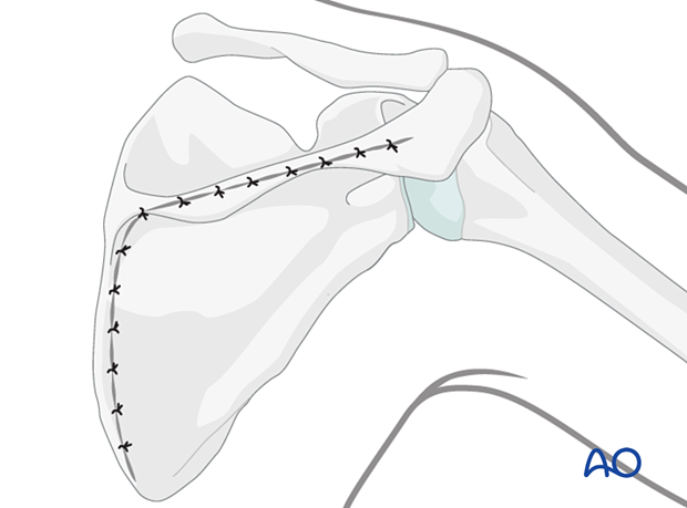
Posterior approach to the scapular body
1. Introduction
Exposure of the body and articular segment of the scapula is based on the extensile Judet approach.
The modified Judet approach avoids detachment of the deltoid muscle and gains almost the same exposure.
This approach permits exposure of the
- lateral column
- articular segment
- medial border and body
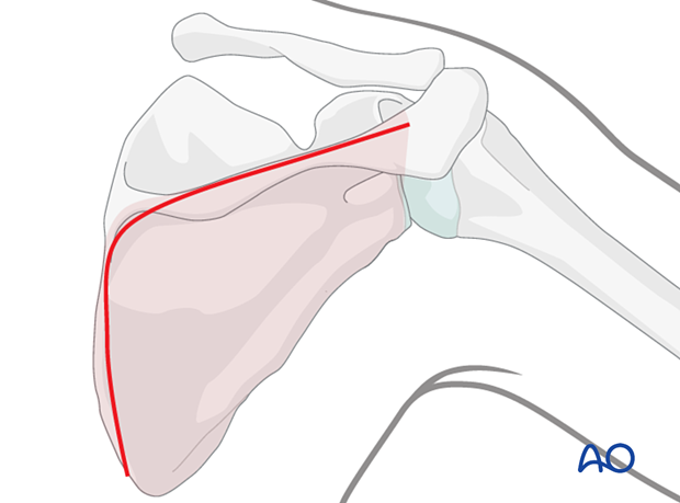
If exposure of the lateral column and art articular segment without exposure of the medial border and body is indicated, a direct lateral column (Brodsky) approach can be performed.
Both exposures are based on an interneural, intervascular concept.
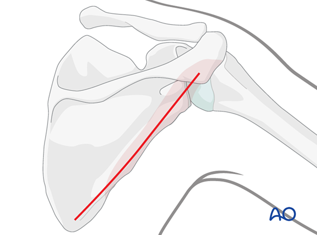
The Brodsky approach may be combined with a limited incision over the medial end of the scapular spine to control a medial border exit fracture line.
Little, if any, muscular detachment is needed for extensile exposure of relevant regions.
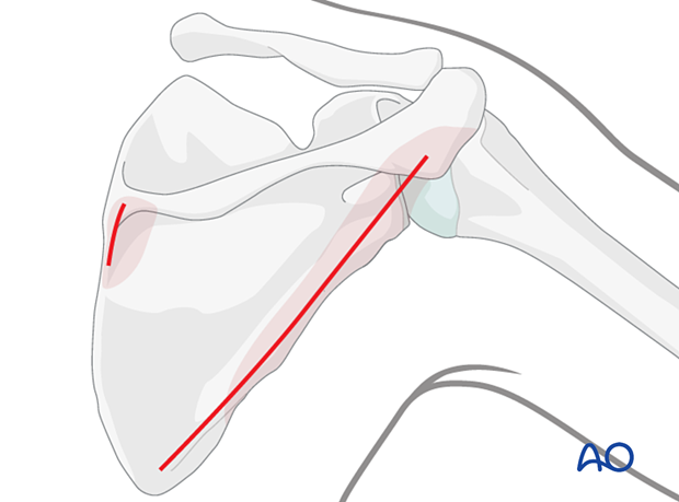
2. Anatomy
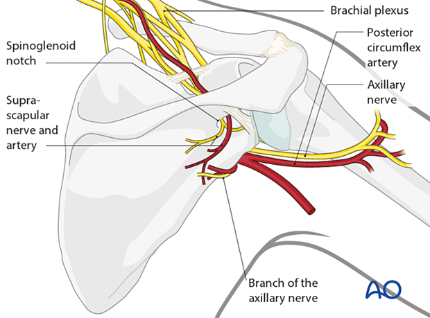
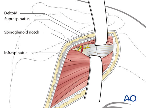
During the posterior approach, care must be taken not to injure the suprascapular nerve at the spinoglenoid notch on its way to supply the infraspinatus muscles.
Teres minor is supplied by a posterior branch of the axillary nerve given off as the nerve emerges from the quadrilateral space with the posterior circumflex artery and vein.
3. Positioning
The lateral decubitus position is preferred.
The injured limb is supported on an arm support with the arm between 45-90° abducted relative to the scapula.
4. Marking of anatomical landmarks
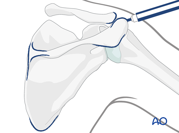
The following anatomical landmarks should be identified and marked:
- Posterior corner of the acromion
- Posterior aspect of the acromioclavicular joint
- Medial end of the scapular spine
- Inferior pole of the scapula
5. Skin incision
Incision for Judet approaches
The incision begins at the posterior corner of the acromion. It continues along the spine of the scapula to its medial border and then distally to the inferior pole of the scapula.
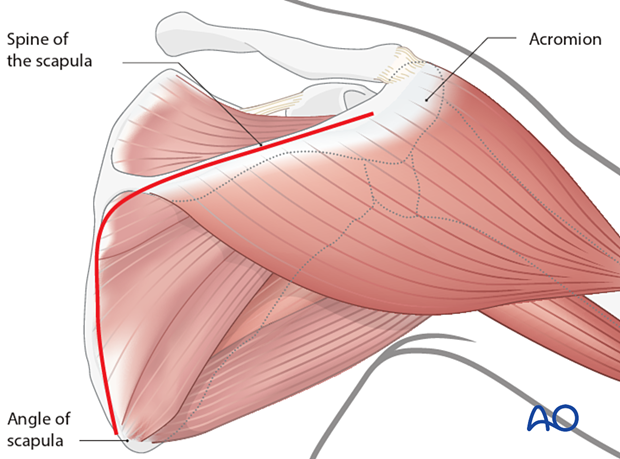
The skin is elevated at the epifascial level, only enough to expose the inferior border of the deltoid muscle.
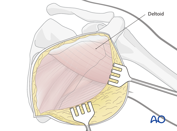
Elevate the of the posterior border of the deltoid to expose the infraspinatus (the modified Judet)
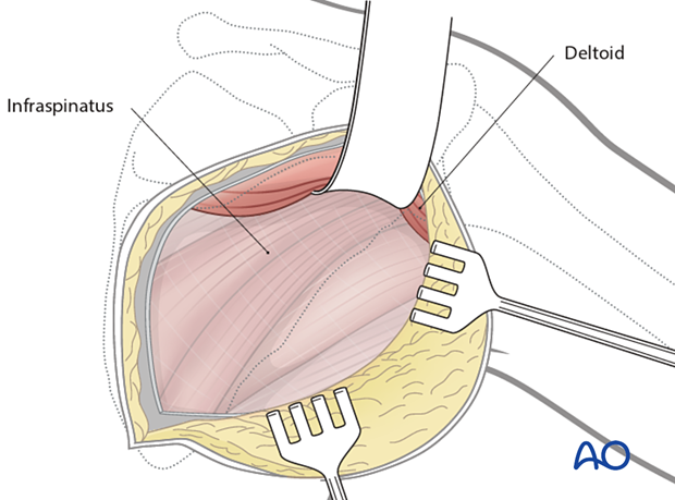
Detach the deltoid from the spine of the scapula for greater access to the superior part of the articular segment if indicated by the fracture pattern (the classic Judet)
Elevate the infraspinatus muscle from the infraspinatus fossa and retract it laterally to expose the entire posterior aspect of the scapula.
This includes:
- the posterior aspect of the neck of the scapula
- posterior glenoid rim
- posterior scapular body
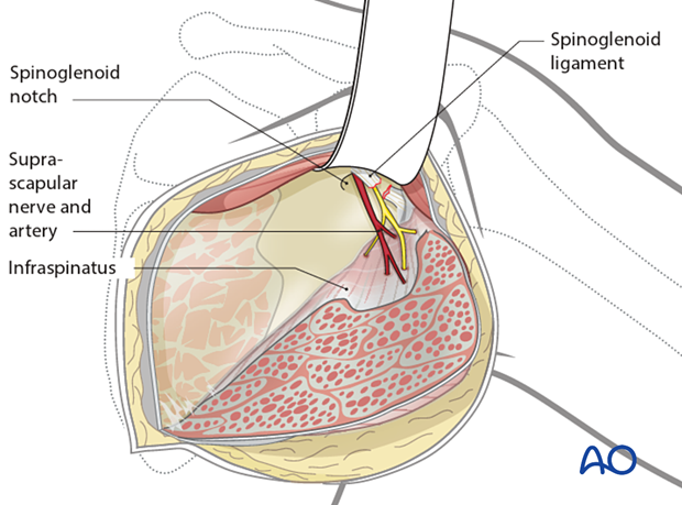
Lateral column approach (Brodsky approach to the scapula)
The incision begins medial to the posterior corner of the acromion and continues distally to the inferior pole of the scapula, parallel to the lateral border of the scapula.
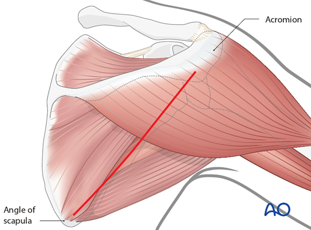
The skin is elevated at the epifascial level enough to expose the inferior border of the deltoid muscle.
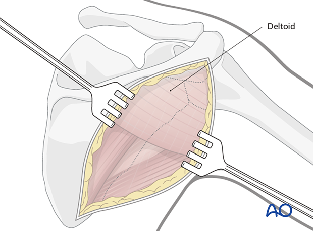
The deltoid muscle is retracted superiorly after incising the fascia over the inferior border of the muscle, taking care to avoid traction on the axillary nerve and posterior circumflex humeral artery laterally. This retraction exposes the infraspinatus fascia, which is incised in the line of the muscular fibers.
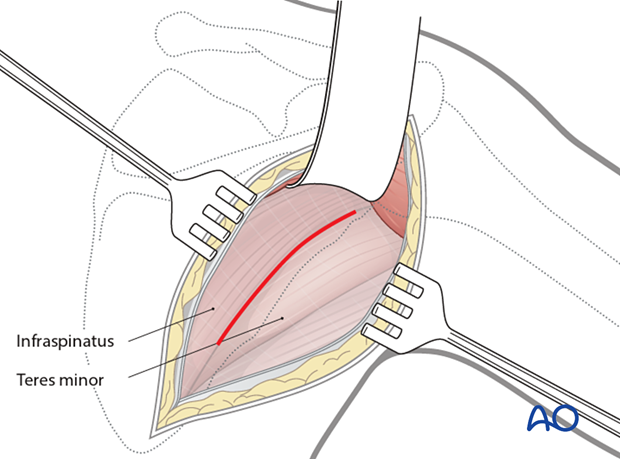
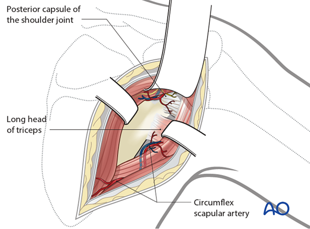
The interval between the infraspinatus and the teres minor is identified. The interval is developed to expose the long head of the triceps laterally. By following this muscle upwards to its attachment to the infraglenoid tubercle, the inferior and posterior capsule of the joint and the inferolateral border of the scapula are exposed.
Exposure of the superior part of the articular segment and glenoid rim can be performed through the interval between the supraspinatus and infraspinatus. Care must be taken to avoid the injury of the nerves to infraspinatus at the spinoglenoid notch.
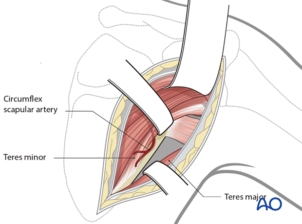
The interval between teres minor and teres major is identified by exposure of the circumflex scapular vessels. This interval permits exposure of the remainder of the lateral column.
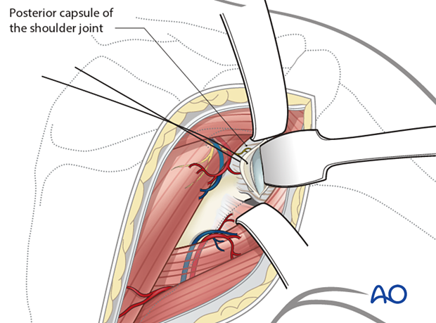
A transverse posterior glenohumeral arthrotomy exposes the glenoid fossa when checking for intra-articular fracture reduction and inadvertent penetration of the joint surfaces by fixation screws.
6. Wound closure
After definitive fixation is completed, the wound is irrigated, and the joint capsule is repaired. The fascia over the infraspinatus muscle is repaired. The deltoid muscle is allowed to fall back into place. It is not necessary to repair the deltoid fascia.
Close the subcutaneous tissues and the skin.
