ORIF - Tension band
1. Introduction
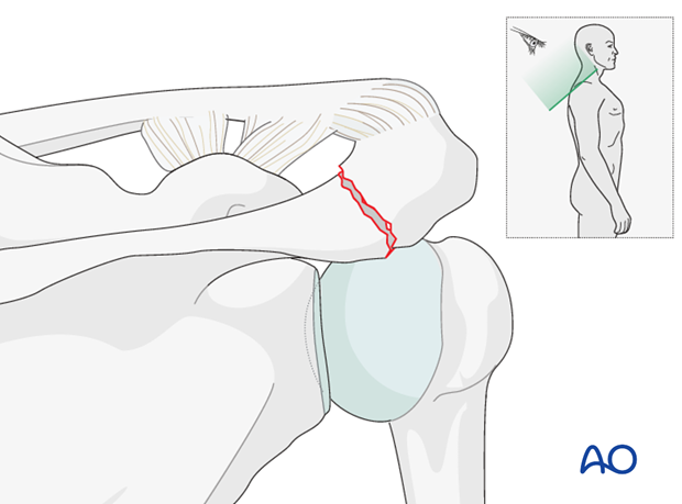
Most isolated acromial factures can be managed non-operatively.
However, typically most acromial fractures are associated with more complex fractures of the scapula, and/or clavicle, and may also involve the suspensory ligament complex.
Any disruption of the ligament complex is treated as described in the section of LSSS.
Most of the acromion is cancellous bone and provides poor fixation for screws.
2. Patient preparation
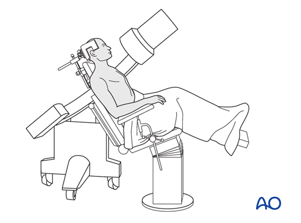
Depending on the approach, this procedure may be performed with the patient in a beach chair position or lateral position.
3. Approach
For simple fractures of the acromion, a superior approach, ie a superior scapular approach or a sabercut approach, is recommended.
For associated complex injuries requiring an anterior approach, the sabercut approach may be extended into the deltopectoral approach (eg, coracoid, glenoid, proximal humerus fractures).
4. Reduction and fixation
Reduction
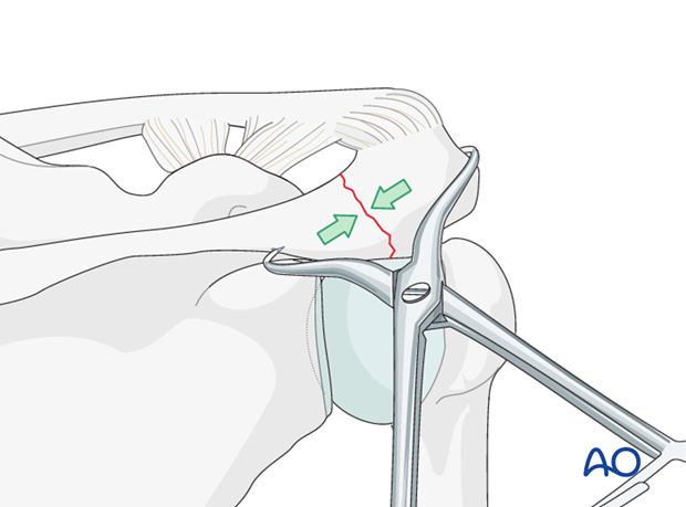
Reduction is best achieved using a reduction clamp.
K-wire insertion
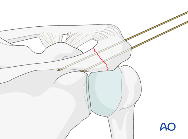
In a simple transverse fracture, a tension band alone may be sufficient.
For a pure tension band, a K-wire is inserted from the lateral side and directed slightly posteriorly so that it exits the spine of the scapula on the other side of the fracture. A second K-wire is inserted parallel to the first with the help of a parallel drill guide. Both K-wires should protrude 5 mm dorsally.
Check the position of the K-wires with an image intensifier to make sure they are not inserted subacromially and have penetrated the rotator cuff or other structures.
Drill with the 2 mm drill bit a transverse hole through the spine of the scapula, at least 2-3 cm distal to the fracture. Then insert the wire through the transverse hole and then around the two K-wires in such a way that you will tighten simultaneously both the loop and the wires. This will achieve symmetrical tension in both wires.
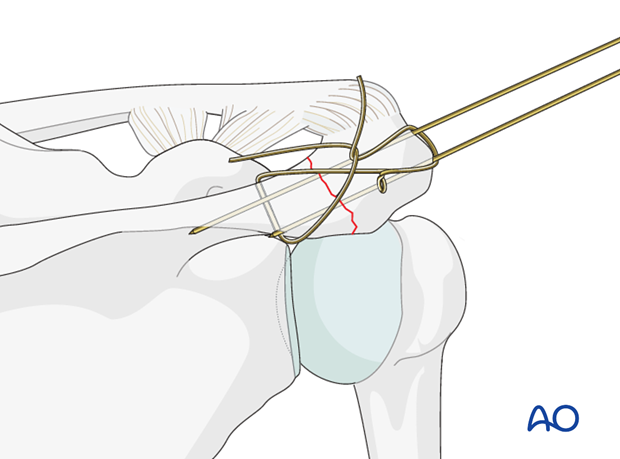
The lateral ends of the K-wires are bent to a 90 degree angle and cut, leaving a 1 cm stump.
This will pull the K-wires 2-3 mm laterally.
The 90 degree angle is then further bent to a 180 degree angle before the "hook" is sunk into the bone. This will restore the 5mm protrusion of the K-wires dorsally.
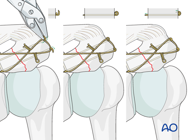
Check the reduction and fixation with an image intensifier.
5. Aftercare
The aftercare can be divided into 4 phases:
- Inflammatory phase (week 1–3)
- Early repair phase (week 4–6)
- Late repair and early tissue remodeling phase (week 7–12)
- Remodeling and reintegration phase (week 13 onwards)
Full details on each phase can be found here.


