Deltopectoral approach to the scapula
1. Introduction
The (anterior) deltopectoral approach can be used for almost any shoulder fracture treatment and is often the preferred approach, especially in anterior glenoid fractures.
This approach is also highly recommended for extensile exposures.
2. Anatomy
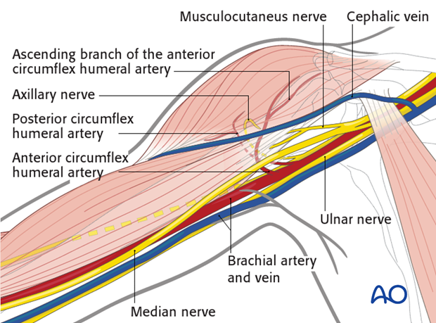
The course of the following neurovascular structures should be kept in mind:
- Cephalic vein
- Anterior circumflex humeral artery
- Ascending arcuate branch of the anterior circumflex humeral artery
- Posterior circumflex humeral artery
- Musculocutaneus nerve
- Axillary nerve
- Suprascapular nerve and artery
Further neurovascular structures, eg, the brachial plexus, are only at risk if there is an excessive retraction.
3. Skin incision
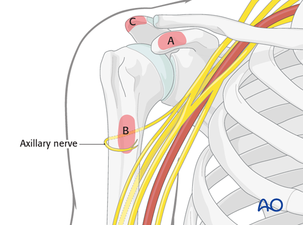
Anatomical landmarks for the anterior deltopectoral approach are:
A) Coracoid process
B) Proximal humeral shaft (on the level of the axilla)
C) Acromion
Sound knowledge of surface anatomy is essential before any skin incision can be made.
Make a 12-14 cm long skin incision between the coracoid process and the proximal humeral shaft towards the deltoid insertion.
A superior extension can be made by extending the incision superiorly to the AC joint or the acromion.
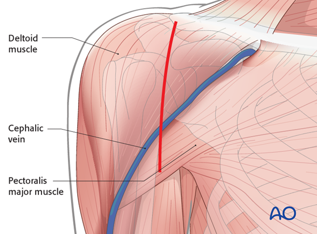
4. Exposure of deltopectoral interval
An interval is developed between the pectoralis major and the deltoid, preserving the cephalic vein which can be taken either laterally or medially.
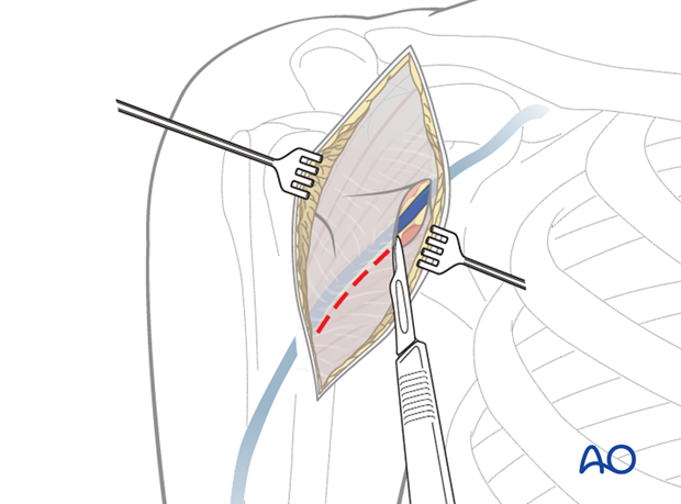
5. Dissection down to the deltopectoral groove
Retract the cephalic vein laterally or medially, and open along the groove. If retracted laterally, the anatomical drainage of blood from the deltoid muscle is respected but it is at risk of damage by retractors during surgery. In any case, the cephalic vein should be preserved.
Failure to find the deltopectoral groove can lead to difficulty in dissection of the deltoid and possibly to denervation of the anterior portion of the deltoid.
Bluntly dissect between and under the deltoid and pectoralis muscles down to expose the clavipectoral fascia.
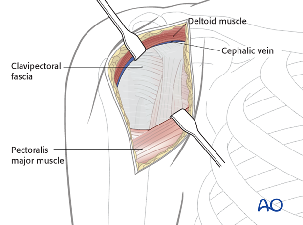
6. Exposure
Identify the coracoid process and the conjoined tendon.
Incise the clavipectoral fascia lateral to the conjoined tendon and inferior the coracoacromial ligament.
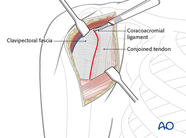
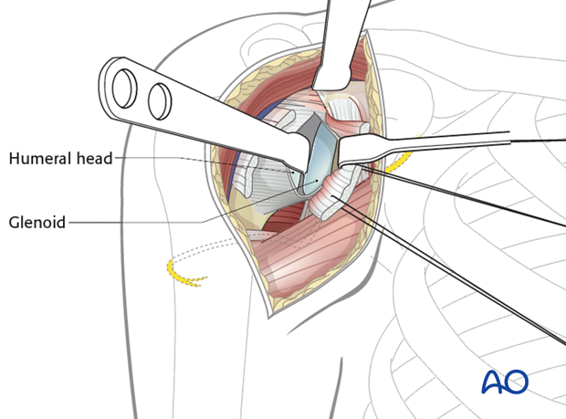
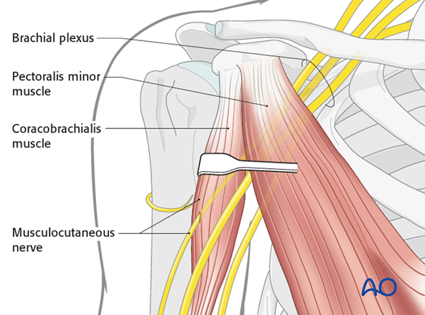
Retract the deltoid muscle laterally using a delta (modified blunt Hohmann) retractor and the conjoint tendon medially using a right angle retractor. The musculocutaneous nerve enters the coracobrachialis muscle as close as 2.5 cm distal to the tip of the coracoid. Retractors placed under the conjoined tendon can cause neurapraxia or even permanent damage to the musculocutaneous nerve; therefore vigorous medial retraction must be avoided.
Take care regarding the musculocutaneous nerve and underlying brachial plexus. Avoid excessive traction medially.
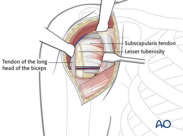
The subscapularis tendon is identified and divided vertically lateral to the musculotendinous junction. Remember the axillary nerve runs superficial to the subscapularis muscle medially before turning inferiorly and posteriorly. It is not seen in this exposure but must be kept in mind!
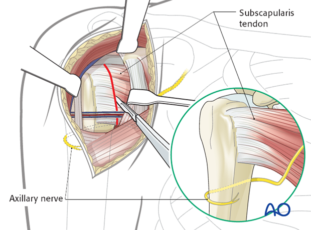
Reflect the subscapularis from the underlying joint capsule and enter the joint through a vertical capsulotomy, medial to the lateral stump of subscapularis.
Do not forget that inferiorly to the glenoid the axillary nerve runs posteriorly to enter the triangular space.

The humeral head is then retracted in order to visualize the glenoid.

7. Closure
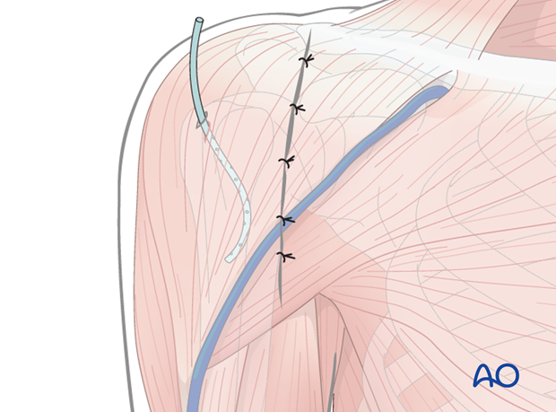
The arthrotomy is repaired by suture closure of the capsule and then the subscapularis.
Irrigate the wound. A drain may be placed if needed.
Meticulous hemostasis should be maintained throughout the procedure. Persistent bleeding usually involves the cephalic vein or its branches, or the trio of inferior vessels on the tendon of the subscapularis.
Close the deltopectoral groove, the subcutaneous tissues and the skin.
