Functional anatomy and biomechanical concepts in the hand
1. Introduction
The hand, complementing visual expression and language as our means of communication and as the tool through which we interact with the environment, comprises simple, repeating units combined as a system of sensate levers bringing the palmar sensory receptors of the fingers into optimal relation with the object of the visual attention.
The hand also functions as a load bearing platform: during power grip (involving the whole hand), and pincer or tripod grip (involving mainly the radial column of the hand), the intracarpal relationships change, guided and limited by the intercarpal ligaments, to distribute the resultant load to the radius and ulna at the distal forearm. This adjustment brings the carpal bones into an energy-efficient interosseous-stable state of ‘close-packing’, permitting load-transfer with minimal effort from the brachio-(meta)carpal muscles.
If a finger becomes functionally isolated by injury or splintage and does not work with the rest of the fingers of the hand, global hand function can be impaired. Stiffness caused by interstitial edema leads to loss of functional movements including loss of grip strength. Additionally, sensory disturbance can make the finger more easily reinjured.
2. The sensate hand
Introduction
The hand is a multifunctional tactile tool to explore the environment comprising a finely interrelated system of repeated units (the digits) and sensory systems. Understanding these sensory systems, particularly protective sensation, tactile discrimination, and discriminative sensation, is crucial for accurate diagnosis and treatment of hand injuries.
Protective sensation
- Function: provides immediate reflex withdrawal from injurious stimuli like heat, cold, and sharp objects
- Neural pathway: mediated by large, myelinated Aβ fibers (therefore fast transmission) traveling in the median, ulnar, and radial nerves
- Clinical evaluation: light touch with a wisp of cotton, testing heat and cold perception (metal probes), pinprick testing (Semmes-Weinstein monofilaments)
- Clinical significance: impaired protective sensation can lead to burns, lacerations, and self-inflicted injuries
Tactile discrimination
- Function: enables fine touch perception, allowing for discrimination of textures, shapes, and object properties
- Neural pathway: mediated by smaller, myelinated Aβ fibers and unmyelinated Aδ fibers (therefore slower transmission) within the median, ulnar, and radial nerves
- Clinical evaluation: two-point discrimination test (using calipers), texture recognition (sandpaper, silk), graphesthesia (identifying letters drawn on palm)
- Clinical significance: crucial for daily activities like manipulating objects, dressing, and fine motor control
Discriminative sensation
- Function: provides precise localization of touch and pressure, essential for skilled hand movements and tool use
- Neural pathway: mediated by specialized low-threshold mechanoreceptors (Meissner’s corpuscles, Pacinian corpuscles) and small myelinated Aβ fibers
- Clinical evaluation: localization of light touch, stereognosis (identifying objects by touch without sight), fingertip localization
- Clinical significance: impacts dexterity and ability to perform complex hand tasks
Neural anatomy
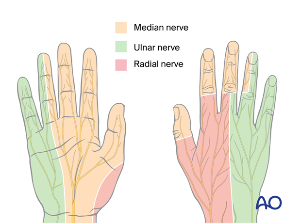
IntroductionThe median, ulnar, and radial nerves provide sensory innervation to the hand.
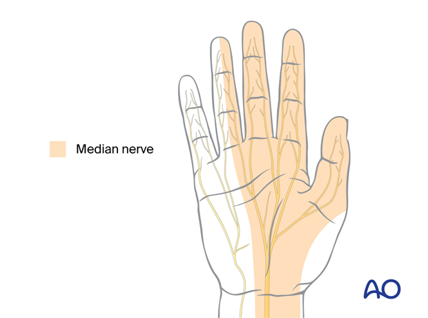
The median nerve branches into the terminal digital nerves to the thumb, index and middle finger as well as the radial side of the ring finger serving the specific function of tripod opposition grip.
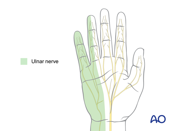
The ulnar nerve branches into the palmar cutaneous nerves and terminal digital nerves of the little and ring finger serving the function of power grip.
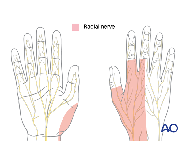
The radial nerve branches into the dorsal digital nerves of all fingers and the back of the hand serving the function of protective sensation.
- Sensory distribution: thumb (excluding palmar aspect of distal phalanx), index finger, middle finger, half of ring finger (palmar and radial aspects)
- Protective sensation: palmar and radial aspects of thumb, index, middle, and half of ring finger
- Tactile discrimination: excellent throughout its distribution, especially fingertips
- Discriminative sensation: excellent, especially fingertips, crucial for fine motor control
- Sensory distribution: little finger, ulnar half of ring finger (palmar and dorsal aspects), palmar aspect of half of middle finger
- Protective sensation: little finger, ulnar half of ring finger, palmar aspect of half of middle finger
- Tactile discrimination: moderate, better on little finger and ulnar half of ring finger
- Discriminative sensation: moderate, important for grip and object manipulation
- Sensory distribution: dorsum of thumb, index finger, middle finger, radial half of ring finger
- Protective sensation: dorsum of thumb, index, middle, and radial half of ring finger
- Tactile discrimination: moderate, better on fingertips
- Discriminative sensation: moderate, important for proprioception and grip control
3. The gripping hand
Extensor tendon mechanism
The extensor tendon mechanism is responsible for extending the fingers at the MCP and interphalangeal joints.
For more details, refer to the chapter on the extensor tendon mechanism.
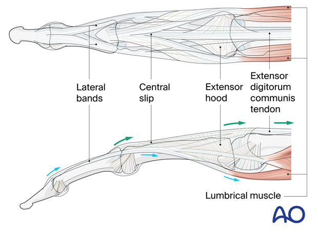
Flexor tendon mechanism
The flexor tendon mechanism is responsible for flexing the fingers at the MCP and interphalangeal joints.
For more details, refer to the chapter on the flexor tendon mechanism.
Tripod/pincer grip
The tripod grip is the foundation for many daily activities. Recognizing its components, potential pathologies, and assessment techniques is crucial to identify and address grip dysfunction effectively. Understanding the tripod grip enhances comprehensive hand care and improves patient outcomes.
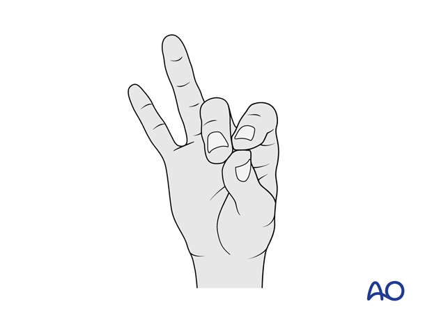
- Thumb opposition: Opposition of the thumb provides the pinching movement and force for the tripod grasp. Fractures and fracture-dislocations may compromise this movement. Therefore, restoration of the anatomy of the joint after injury is essential.
- Metacarpophalangeal (MCP) flexion: Flexion of the thumb and index MCP joints creates the “arch” of the tripod. Fractures and fracture-dislocations which result in malrotation may compromise this arch. Therefore, attention to rotational alignment is essential.
- Interphalangeal (IP) flexion: Coordinated movement at the IP joints provides for fine-tuning of grip strength and object manipulation. Fractures and fracture-dislocations which result in rotational or translational malalignment may compromise this coordination. Therefore, attention to the prevention of malalignment is essential.
- Intrinsic muscle coordination: The thenar muscles abduct, flex, and rotate the thumb to bring it into opposition to the flexing fingers. The fine control of finger flexion requires activation of the interossei and lumbrical muscles.
4. The weight bearing hand
Power grip
The power grip requires forceful flexion of all four fingers with the thumb in opposition for optimal stability. Power is enhanced by dorsiflexion of the wrist.
- Muscles involved: The common flexor muscles of the forearm produce primary flexion power in the thumb and fingers. The common extensor muscles of the wrist and fingers produces dorsiflexion to optimize the flexor biomechanics. Fine control of grip is the product of coordinated action of the small thenar and hypothenar muscles.
- Biomechanics: The thumb acts as a post, with the digital interossei and lumbricals adducting the fingers and flexing the MCP joints to create a secure clamp.
- Clinical significance: Fractures and fracture-dislocation of the thumb may compromise its function as an opposable post. Fractures and fracture-dislocations of the metacarpals and MCP joints may compromise the formation of the stable arch and prevent effective closure of the clamp.
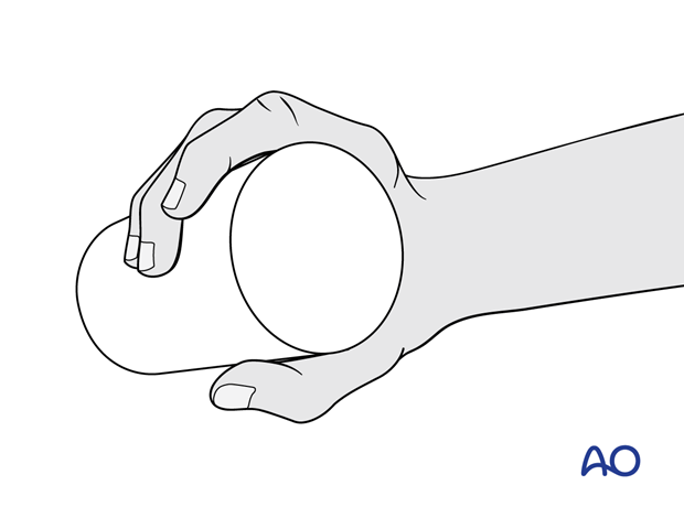
During a power grip, all extrinsic tendons are under tension. This results in rotation and translation of the carpal bones and carpal rows such that there is optimal interosseous stability. This relationship is defined as the closed packed orientation of the carpus.
The intrinsic carpal stability now requires less extrinsic muscular force to maintain power grip. It is therefore an optimal energy efficient posture of the hand for power grip.
Clinical significance: For example, scapholunate ligament rupture precludes optimal close packing of the proximal carpal row. As a result, it is impossible to generate a power grip. Therefore, repair and reconstruction of carpal ligament ruptures are essential for optimal hand function.
Palmar weight bearing
When the wrist is dorsiflexed and the hand open, eg, when falling to take weight to the hand, the carpus is not close packed. It is intrinsically relatively less stable.
Clinical significance: A fall to the outstretched hand is often a cause for distal radial and ulnar fractures. Fracture-dislocations and ligament ruptures of the carpus are frequently associated with distal forearm fractures due to the failure of carpal close packing.
5. References
- Brand PW. Biomechanics of balance in the hand. J Hand Ther. 1993 Oct–Dec;6(4):247–251.
- Brown HG. Functional anatomy of the hand. Orthop Clin North Am. 1970 Nov;1(2):199–204.
- Duncan SFM, Saracevic CE, Kakinoki R. Biomechanics of the hand. Hand Clin. 2013 Nov;29(4):483–492.
- Hirt B, Seyhan H, Wagner M, et al. Hand and Wrist Anatomy and Biomechanics: A Comprehensive Guide. 1st ed. Stuttgart: Thieme Medical Publishers; 2016.
- Imaeda T, An KN, Cooney 3rd WP. Functional anatomy and biomechanics of the thumb. Hand Clin. 1992 Feb;8(1):9–15.
- Moran SL, Berger RA. Biomechanics and hand trauma: what you need. Hand Clin. 2003 Feb;19(1):17–31.
- Pei D, Olikkal P, Adali T, et al. Dynamical Synergies of Multidigit Hand Prehension. Sensors (Basel). 2022 May 31;22(11):4177.


