Anterior approach (Henry) to the forearm shaft
1. Introduction
The anterior (Henry) approach offers good exposure of the whole length of the radius. The length of the incision depends on the extent of exposure needed. The Henry approach in the proximal forearm might result in a more obvious scar.
The landmarks for the skin incision are:
- Proximally: the biceps tendon which crosses the front of the elbow joint, medial to the brachioradialis muscle. The distal landmark is the radial styloid process.
- The brachioradialis muscle is part of the mobile wad, comprising the brachioradialis and the extensors carpi radialis brevis and longus.
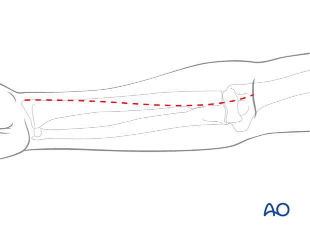
This illustration shows the extents of the incisions for the anterior approaches to the
- proximal third
- middle third
- distal third
of the radial shaft.
Note: The posterolateral (Thompson) approach also offers good access to either the middle or distal third of the radial shaft.
2. Superficial dissection
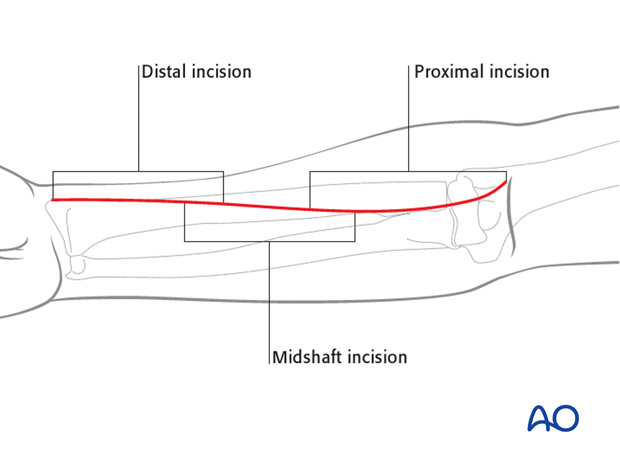
The superficial muscular dissection is similar for all three parts of the Henry anterior approach, illustrated here for the proximal third.
Develop the interval between the brachioradialis (mobile wad) and flexor carpi radialis muscles. The radial artery lies deep to the brachioradialis in the middle part of the forearm and between the tendons of brachioradialis and flexor carpi radialis distally. It can be identified by its two venae comitantes, which run alongside it.

The arterial branches arising from the lateral side of the radial artery are identified by slipping a finger underneath them, lateral to the artery. These are largely the recurrent branches and those to the “mobile wad”.
These branches should be ligated to retract the artery medially.
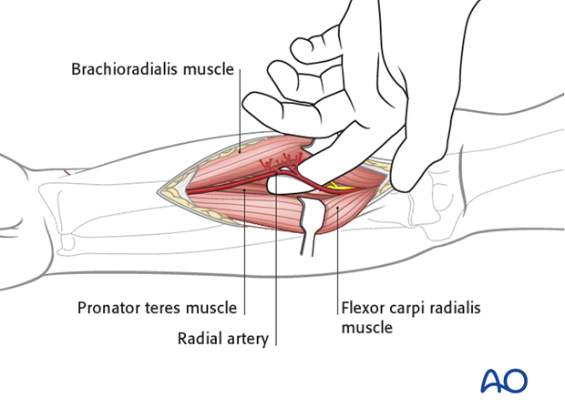
The superficial radial nerve runs under the brachioradialis muscle on the lateral aspect of the radial artery and should be retracted laterally.
3. Deep dissection: proximal third
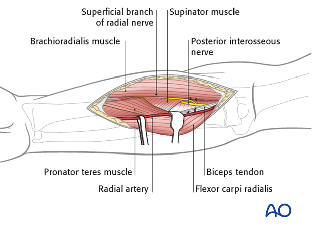
The supinator muscle covers the lateral aspect of the proximal radius. The posterior interosseous nerve (also known as deep branch of the radial nerve) lies within the substance of the supinator muscle. The forearm should be fully supinated to displace the posterior interosseous nerve away from the surgical field.
The supinator muscle is then incised along its most medial edge, and gently elevated from the bone surface only to the extent that it is necessary for the required exposure. The plate is inserted deep to the elevated supinator muscle.
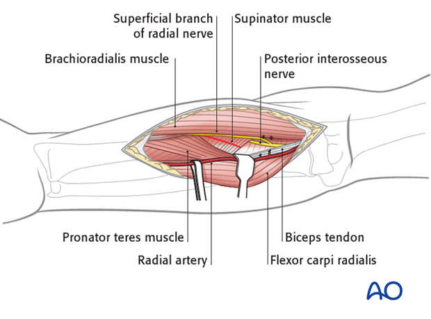
Note: Make sure that the plate is seated on the bone without any soft-tissue interposition. In rare cases, the posterior interosseous nerve itself is identified. In such cases it is recommended that the position of the nerve in relationship to the plate be noted and later documented in the operation report.
4. Deep dissection: middle third
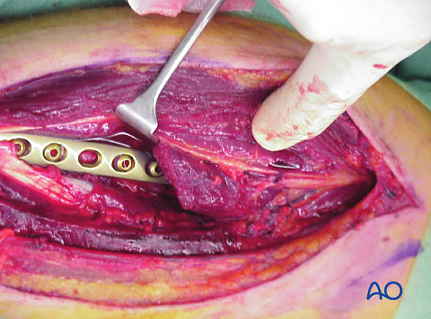
The forearm should be fully pronated to expose the lateral border of the pronator teres muscle and its insertion.
Note: Sometimes it will be necessary to partially detach the pronator teres from the radius. Whenever possible, preserve at least some of its insertion.
5. Deep dissection: distal third (classical Henry approach)
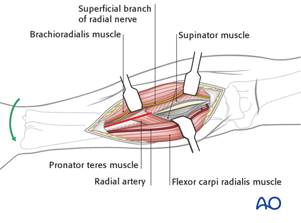
Pronating the forearm will expose the aspect of the radius just lateral to the edge of the flexor carpi radialis, deep to which lie flexor pollicis longus and the pronator quadratus, which become visible when the forearm is supinated.

The forearm is again supinated, and the exposure of the bone is completed by any necessary elevation of the flexor pollicis longus and, more distally, pronator quadratus.

6. Deep dissection: distal third (modified Henry approach)
Note: where exposure is likely to be confined to the distal radius only, the following modified Henry approach is preferred by some surgeons.
The modified Henry approach utilizes the interval between flexor carpi radialis tendon and the radial artery, whereas the classical Henry approach goes between brachioradialis and the radial artery. The modified approach is medial to the radial artery.
Note: The radial artery and the palmar branch of the median nerve are vulnerable during this approach.

The radial artery is retracted laterally and the flexor carpi radialis is retracted in a medial direction. The pronator quadratus muscle is then exposed by retracting medially the muscle belly of the flexor pollicis longus.
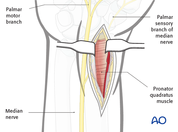
Exposure of the bone is completed by incision of the lateral and distal edges of pronator quadratus muscle leaving a small lateral cuff on the radius to allow for subsequent repair.
This now allows elevation of the muscle belly from the anterior aspect of the distal radius.
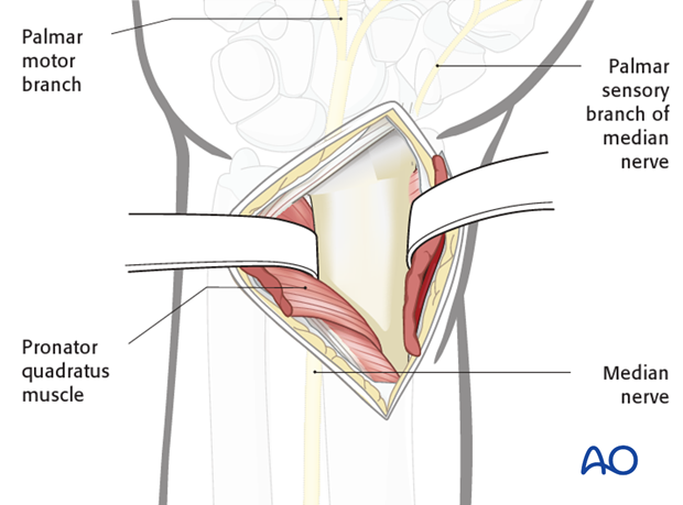
Apart from possible reattachment of pronator quadratus, the anterior deep tissues are left unrepaired. Some subcutaneous sutures are inserted to relieve tension on the skin closure. Wound drainage, either with a closed or an open system, is used. Skin closure is accomplished using interrupted or running sutures, or skin staples.
In certain instances, for example with marked forearm swelling, the wound may have to be left open. There are different techniques to overcome such difficulties (eg, elastic closure, vacuum assisted closure, petroleum jelly gauze, skin substitute, etc).
