Posterolateral approach (Thompson) to the forearm shaft
1. Introduction
The posterolateral (Thompson) approach offers good exposure of the middle and distal thirds of the radial shaft. The skin incision lies straight down the dorsal aspect of the forearm and its length depends on the exposure needed.
The landmarks for skin incision are:
- Proximally: the lateral epicondyle (note that the skin incision will not extend to the lateral epicondyle)
- Distally: Lister’s tubercle
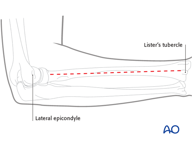
This illustration shows the extent of the posterolateral approach to the middle third of the radial shaft with an extension to its distal third.
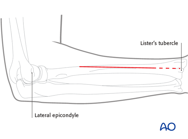
2. Skin incision
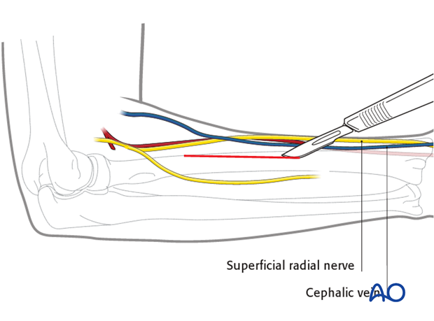
Incise the skin and subcutaneous tissues. In the distal part of the incision, be aware of the superficial radial nerve and the cephalic vein.
3. Deep dissection – middle third
The distal starting point of the deep dissection is the proximal border of the oblique crossing of the abductor pollicis longus muscle over the dorsal surface of the radius. Development of the plane deep to the abductor pollicis longus allows the belly to be lifted with a retractor to give access to the underlying radius.
Continue the dissection proximally between the extensor carpi radialis brevis, which should be retracted laterally, and the extensor digitorum communis which should be retracted medially.
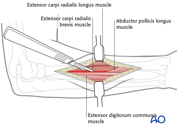
Exposure of the supinator and abductors pollicis
The retraction of the extensor carpi radialis brevis and the extensor digitorum communis will expose…
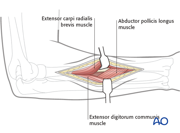
… the supinator proximally and the rest of the abductor pollicis longus distally.
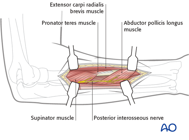
Turn the arm
Turn the arm in full supination to bring the origin of the supinator into view and to move the posterior interosseous nerve away from the area of dissection.
Some elevation of the supinator attachment to the radius is usually necessary to expose the underlying shaft.
If it is necessary to mobilize the pronator teres, should the fracture configuration demand, it can be partially divided at its insertion into the radius, leaving sufficient tendinous cuff attached to the radius to aid subsequent repair.
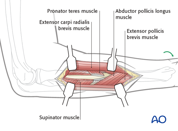
4. Deep dissection – extension to proximal third
Note: Proximal extension of the exposure of the middle third carries considerable risk unless great care is taken to avoid injury of the posterior interosseous nerve.
Identify the posterior interosseous nerve
The supinator wraps around the upper part of the radius. The posterior interosseous nerve runs within its substance. The nerve must be identified and protected as it traverses the muscle. To verify the position of the posterior interosseous nerve, a small hole can be made into the supinator muscle. The nerve can also be felt as a bulge in the muscle.
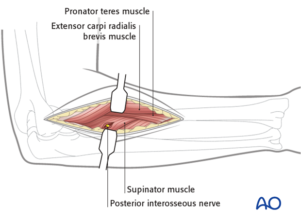
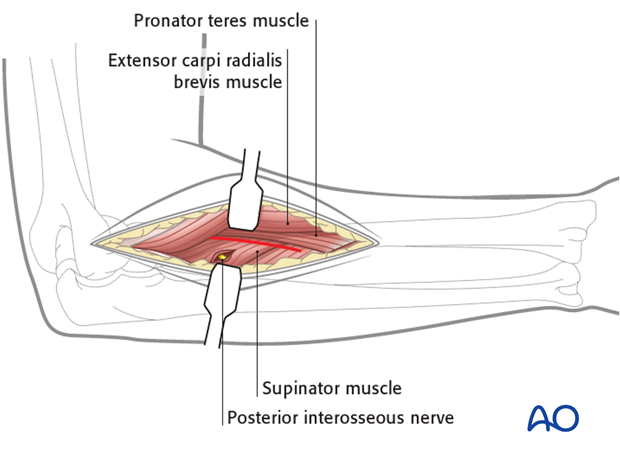
The elevation of the supinator involves an incision along its insertion into the radius. This may be aided by a degree of supination of the forearm.
The supinated muscle should be elevated subperiosteally, to protect the posterior interosseous nerve.
5. Distal extension
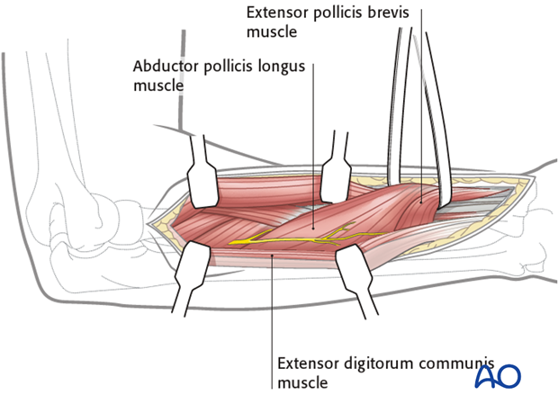
Extend the skin incision to the Lister’s tubercle. Undermine the abductor pollicis longus and extensor pollicis brevis and lift them up with a vessel loop. Towards the Lister’s tubercle, retract the tendon of the extensor carpi radialis longus laterally. Distally, a partial incision of the extensor retinaculum is often necessary: respect the extensor pollicis longus tendon on the medial side of the tubercle.
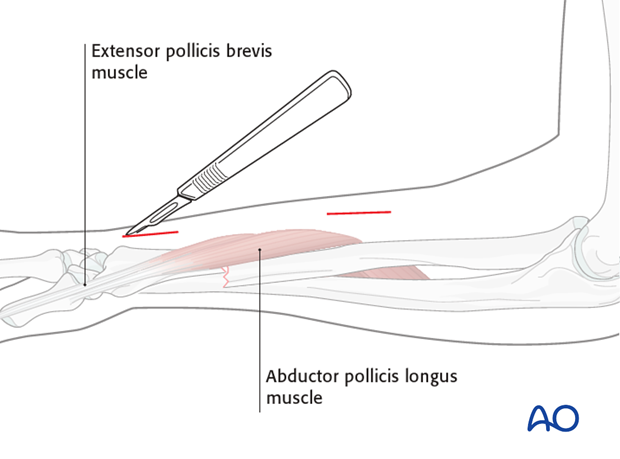
6. Approach for minimally invasive techniques
An option to the described open approach is the minimally invasive approach, making two small incisions proximal and distal to the abductor pollicis longus muscle.
This approach is described with the detailed procedure.
