Observation
1. Indication
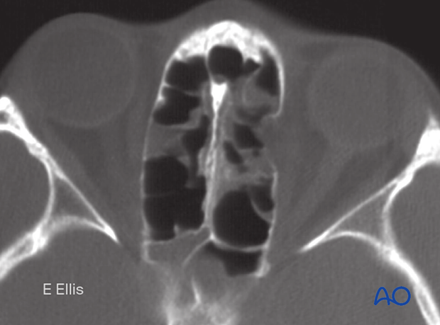
Observation may be indicated in non- or slightly displaced medial orbital wall fractures where an increase in orbital volume is minimal, and there is no continuing disturbance of eye motility.
Observation may also be justified in cases where the patient's condition does not allow for surgical intervention or when the patient is not willing to undergo surgical repair of the fracture.
The decision to observe or to perform surgery is based on thorough clinical and radiographic (CT) evaluation because correction of a potential secondary deformity is challenging.

2. Follow-up
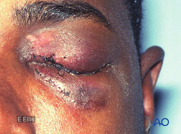
Due to periorbital edema following trauma, the majority of patients may initially present with some degree of proptosis. After resolution of the swelling the patient is reevaluated for enophthalmos or dystopia. Therefore, a decision for operative intervention must be delayed until the swelling has resolved. Furthermore, significant swelling of the soft tissue of the orbit may make surgical intervention more challenging.
The patient must be examined and reassessed regularly.
Additionally, an ophthalmological examination is indicated. If any disturbance of eye mobility or globe position develops, CT re-examination may be indicated, and operative treatment becomes necessary. MRI examination is occasionally useful for additional soft tissue evaluation.

3. Aftercare
For aftercare and rehabilitation following observation, please refer to your local protocol.


