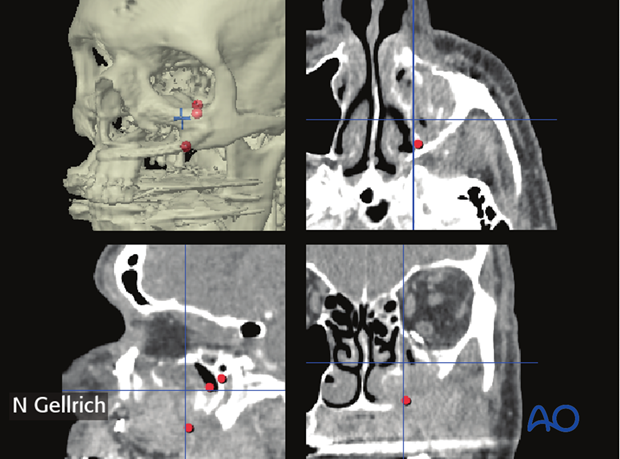
Biopsy mapping
1. Introduction
Indications
Biopsy mapping is used when it is helpful to accurately record the location of biopsy sites.
In ablative surgery of malignancies located in the maxilla or close to the skull base, a complete resection may be challenging due to the complex anatomy including vital structures.
Biopsies including frozen sections are commonly taken at locations where incomplete resections are suspected. The surgeon will typically name the location and it is recorded by a non-sterile assistant. However, no matter how differentiated the naming for each biopsy is, it is very difficult to recall at a later stage the pricewise 3D location. If any of the frozen sections are tumor positive, and no further surgical treatment is indicated, patients may be submitted to postoperative radiotherapy. The planning of radiotherapy is then based on the intraoperative notes describing where the positive biopsies were taken. This process may however be prone to errors due to the 3D complexity and to a possible lack of accuracy in the noted description.
An exact language-independent digital mapping may contribute to a more precise follow up with restaging 3D imaging procedures.
When hard tissues and adjacent soft tissues are involved, a more accurate process can be followed to ensure a correct recording of the sampling locations in all three dimensions.
Here a digital workflow utilizing computer navigation together with appropriate software will be presented. The case used is the treatment of a 64 year old male with recurrence of a squamous cell carcinoma in the left maxillary sinus.
Useful additional reading
2. Imaging
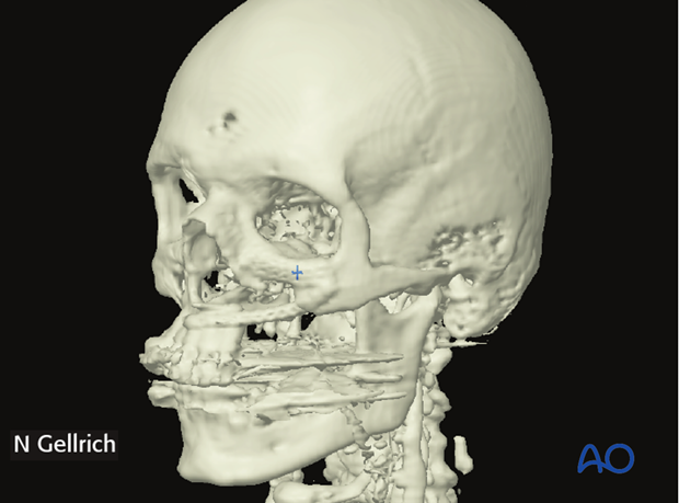
3D-CT scans and/or MRI is/are obtained using navigation fiduciary markers.
In this case, a dental splint attached to the maxilla containing radio opaque markers was used.
Tumor segmentation
There is currently no software available for complete auto segmentation of the tumor. The tumor is therefore manually segmented in the images.
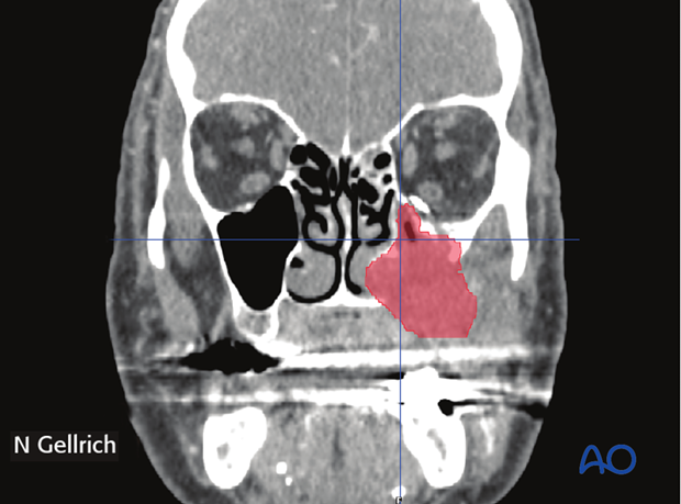
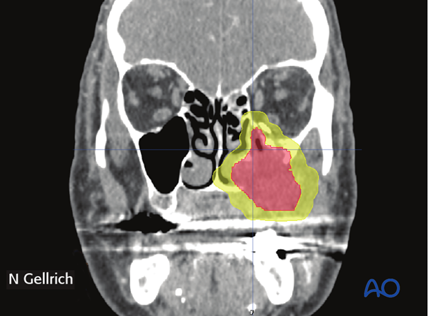
The volume of the tumor (red) is virtually augmented to simulate the size of the required resection including safety margins (yellow). The size of these margins (safety margins) will depend on the tumor histology.
3. Biopsy mapping
The tumor resection is completed and frozen sections are taken from the areas/spots where incomplete resection is suspected. The sampling sites are recorded in the navigation system using the pointer (numbered dots).
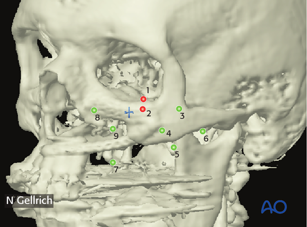
Tumor positive sampling sites are highlighted (red) and the radiotherapist can import the voxel based information directly into his/her radiotherapy planner.
This technique provides the radiotherapist with an exact location of where to focus the radiotherapy.
