Plate fixation
1. Principles
Open fractures
Fractures of the interdental space and horizontal ramus are almost always open fractures. Wound debridement and flushing is extremely important. Nevertheless, infections of soft tissues as well as the bone are common. Therefore, ventral drainage is important.
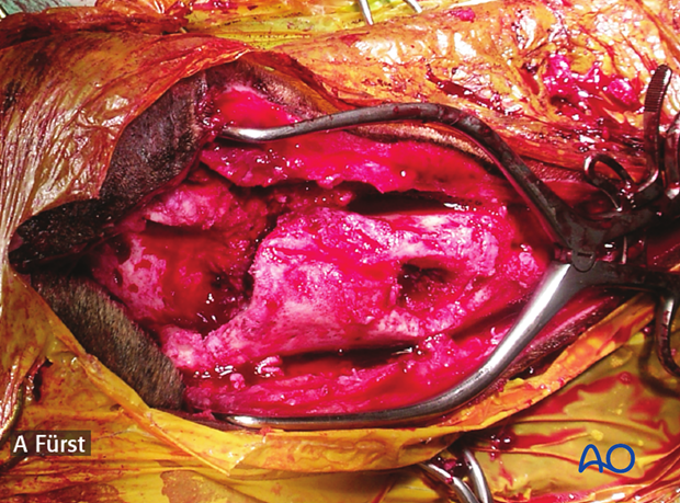
Plate position
Although the oral side of the mandible and maxilla are the tension surfaces, plates are frequently applied ventral to the mandible, where the thick cortex provides a stable fixation.
Some surgeons prefer to place the plate on the lateral side of the mandible. Implant failure is less likely in this location.
Intraoperative imaging
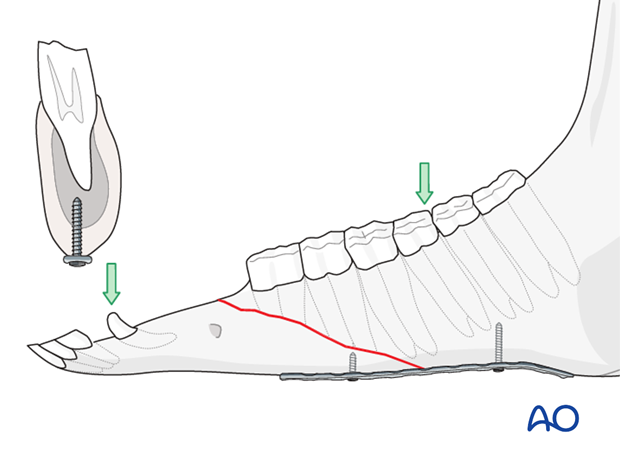
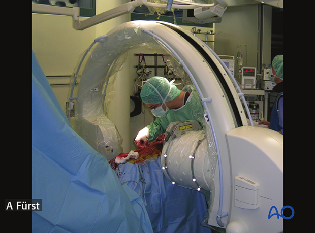
The use of intraoperative imaging is highly recommended to check correct placement of the screws and avoid damage to the tooth roots.
2. Preparation and approach
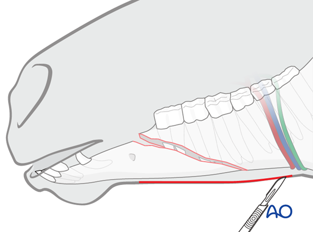
This procedure is performed with the patient placed in dorsal recumbency through the submandibular approach.
3. Reduction and fixation
Plate selection and preparation
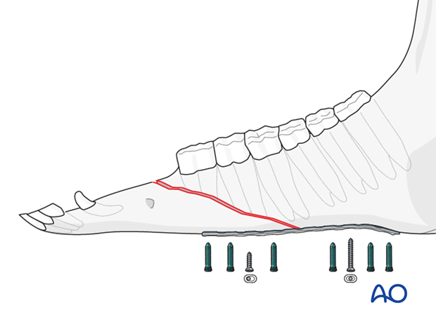
Dynamic compression plates can be used but locking compression plates (LCP) are preferred, because they provide better stability. In areas with only one cortex and a predominance of spongy bone, locking head screws provide superior stability.
Depending on the size of the horse, narrow 3.5 or 4.5 LCPs are used. Usually, 8-12 hole plates are appropriate.
Screw length must be determined precisely to avoid engagement of the tooth roots.
Only minimal plate contouring is required.
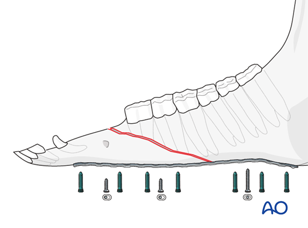
Alternatively, a very long plate (14-16 hole) can be used. In this case, not every hole needs to be filled with a screw.
Wound debridement
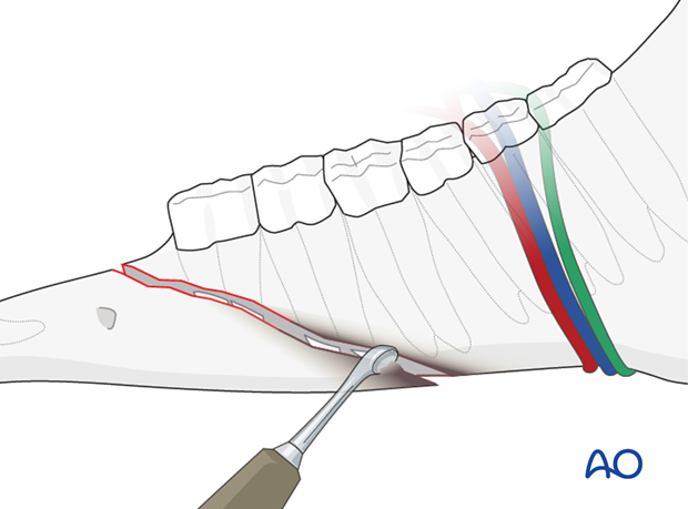
In older or infected fractures, wound debridement is important. All infected bone and tissue must be removed with a curette or rongeur.
Additional flushing is indicated.
Reduction
The fracture is stabilized using pointed reduction forceps.
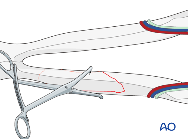
In selected cases interfragmentary screws are applied to maintain reduction and provide compression at the fracture site.
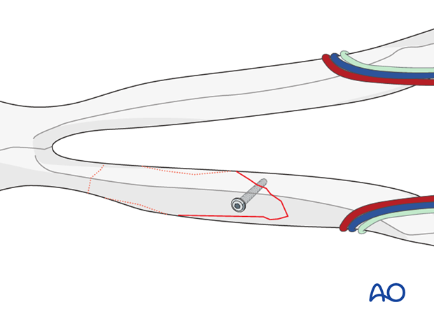
Plate fixation
The plate is placed ventral to the mandible and fixed to the bone with one cortex screw on each side of the fracture.
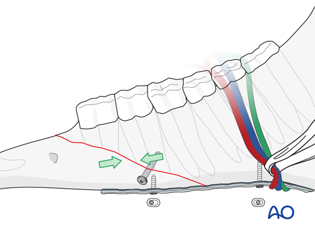
If the plate is to be placed further caudally, on the incisura vasorum, the salivary duct and the blood vessels need to be protected and elevated using a penrose drain. The plate is slid under the bundle onto the bone.
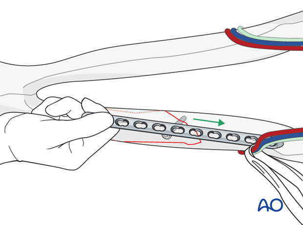
The remaining plate holes are filled with locking screws.
The locking head screws provide good stability, because only one cortex of the mandible can be used for fixation.
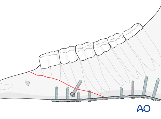
Closure
The skin is closed routinely. In old, infected or open fractures, a drainage should be placed through an additional small incision that is left open.
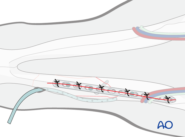
4. Intraoral wire fixation
To increase stability, intraoral wire fixation is applied in addition to the plate. For details on wire fixation see "Wire fixation".
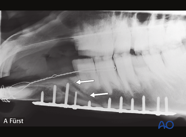
5. Aftercare
During the post-surgical period, the horse is confined to stall rest for one week and fed with soft fiber feed stuff; firm feed such as hay cubes, apples or carrots should not be fed. Antibiotics and anti-inflammatory drugs are given for 3 to 5 days or longer, if required, especially in open fractures. Intraoral cerclage and tension wires are cleaned once daily until their removal. Wound opening is cleaned and flushed daily with antiseptic solutions.
After the fracture has healed plate and wire can be removed under sedation and local anesthesia. It is recommended to remove the plate first and the wires three weeks later to reduce the risk of refracturing.


