

Lag screw and K-wire
1. Principles
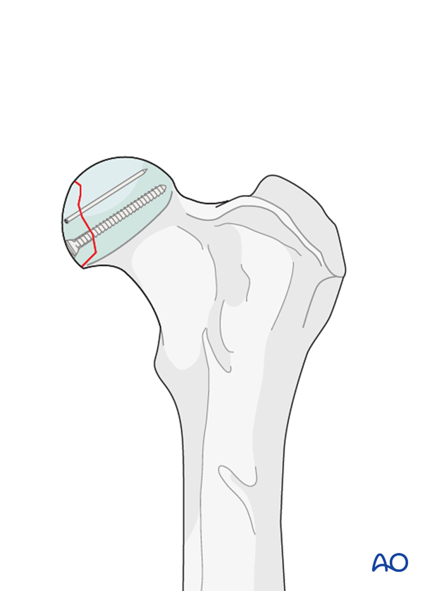
The preferred treatment for 31-C1 fractures is internal fixation with a countersunk screw and an antirotational K-wire. Anatomical reconstruction is required to avoid secondary degenerative changes. Implants applied from the articular surface must be countersunk.
2. Patient positioning
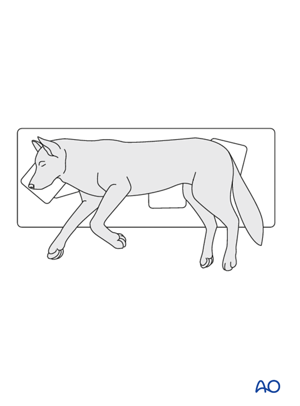
This procedure is performed with the patient in either lateral recumbency....
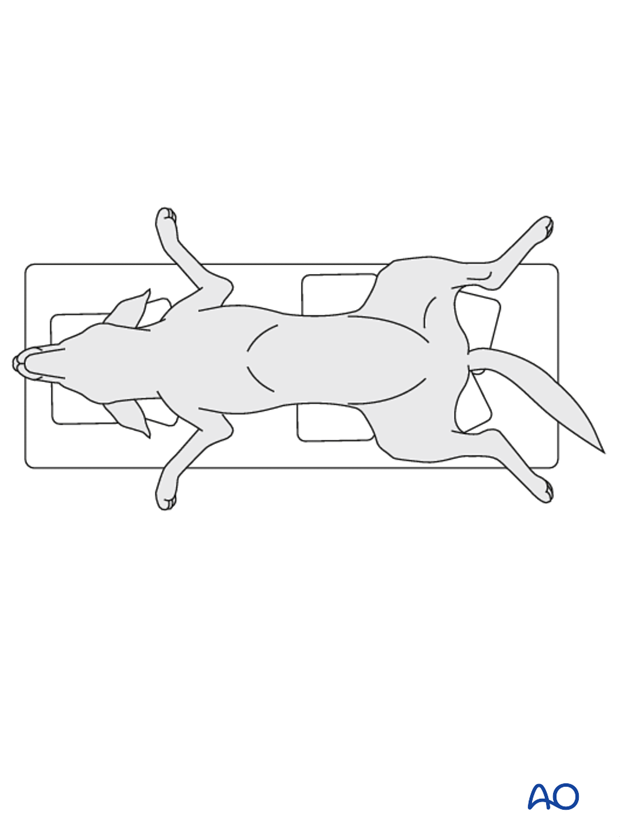
... or dorsal recumbency
3. Approach
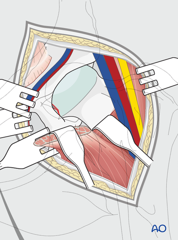
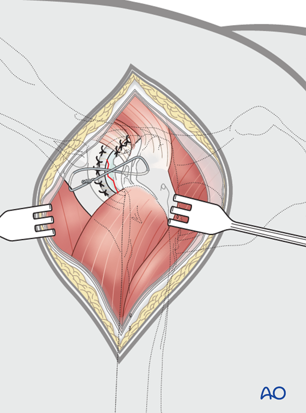
This fracture may be exposed using a craniolateral approach. If further visualization is needed the approach is combined with a dorsal approach. Transection of the ligament of the femoral head may be needed to allow reduction and fixation of the fragment.
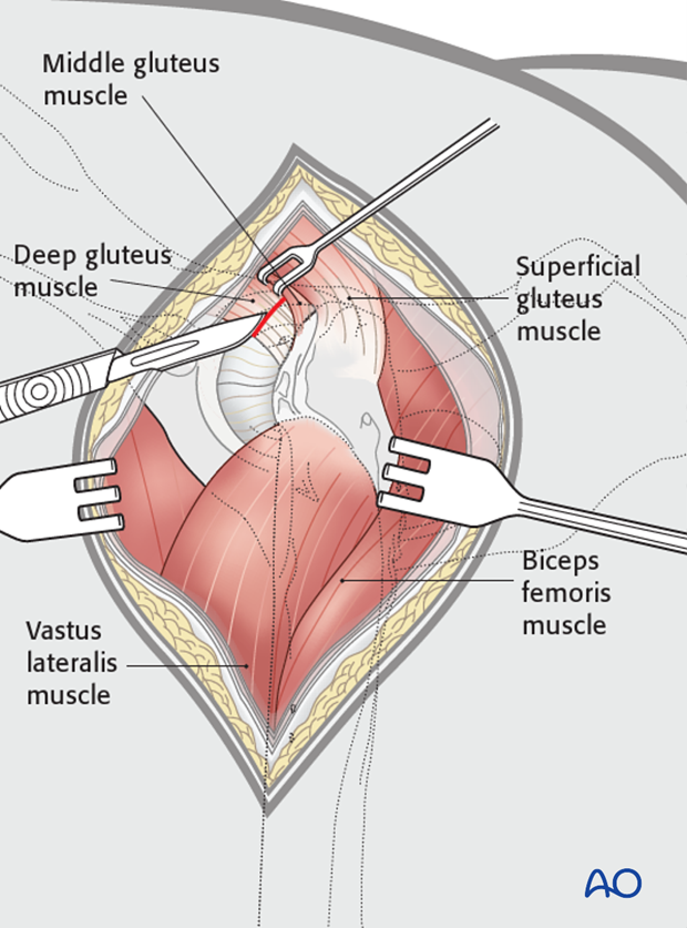
Alternatively, a ventral approach can be used. This approach has the advantage of avoiding the transection of the ligament of the femoral head.
4. Surgical technique
Implant selection
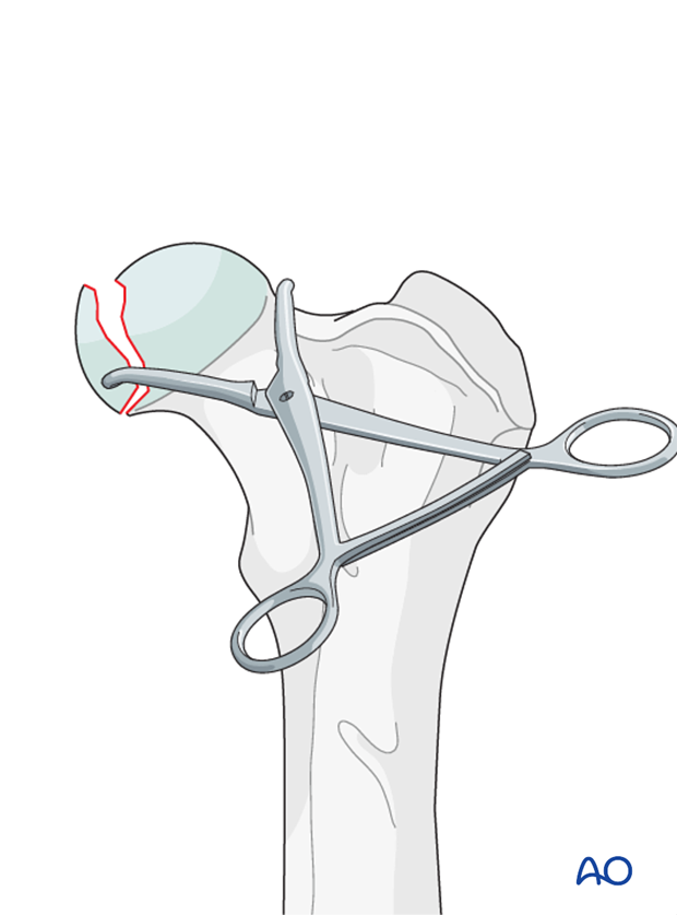
A cortical screw applied in lag fashion combined with an antirotational K-wire is used. In large fragments, two lag screws can be used.
Reduction
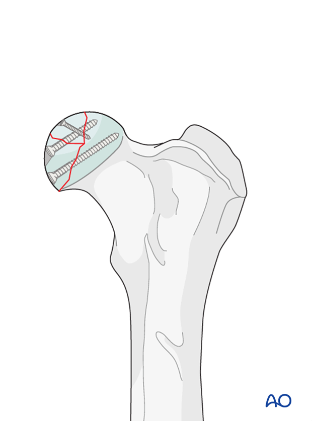
The fracture is reduced and secured with pointed reduction forceps.
Fixation
Lag screws or K-wires, depending on fragment size, are used to stabilize the fracture.
A gliding hole is drilled through the cis or near fragment. The drill sleeve is inserted into the gliding hole and the thread hole is drilled.
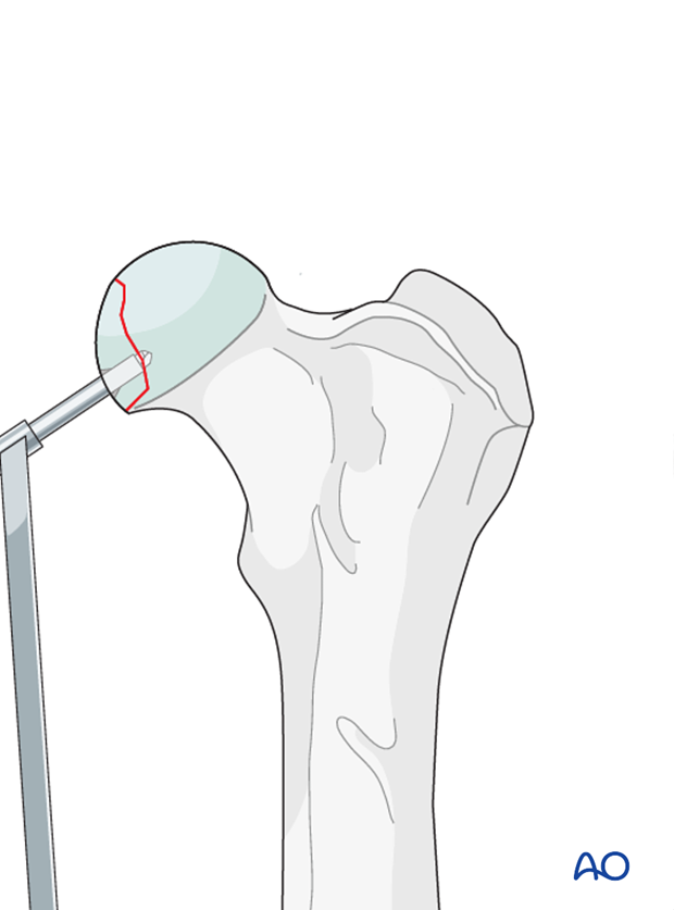
A countersink is used carefully in the glide hole to prepare the bone to accept the screw head. The hole is measured, the thread hole is tapped, and an appropriate length screw inserted.
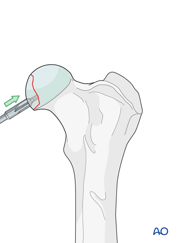
If only one screw was used, an antirotational K-wire is applied and countersunk under the articular cartilage.
Note: Care must be taken to ensure that no implants are protruding in the articular surface.
Read more about lag screw fixation.
Closure
Capsulorrhaphy is performed. If the ligament of the femoral head was transected, additional stabilization of the hip joint is generally required.
See other resources for surgical techniques for traumatic hip luxations.
Validation of fixation
Postoperative orthogonal radiographs are taken to assess fixation.
5. Case example C1
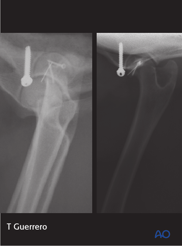
6-year-old Border Collie with a C1 fracture from being hit by a car.

The fracture was repaired using a 1.3 mm lag screw and 2 K-wires countersunk in the articular cartilage. Reduction and stabilization of the hip was achieved with an iliofemoral sling.
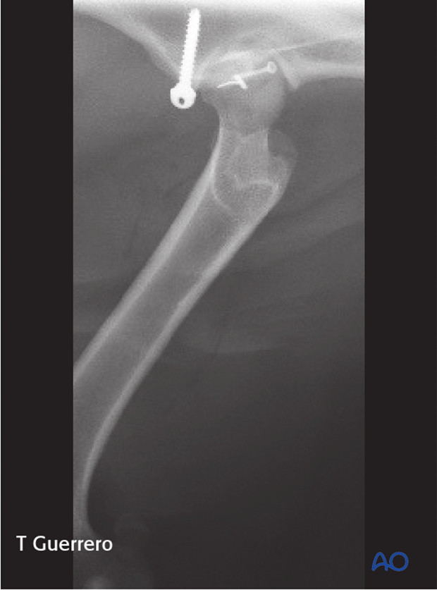
Postoperative radiographs at 6 weeks show complete healing of the fracture.
6. Aftercare
Activity restriction is indicated until evidence of bone union is detected on radiographic examinations.
Implants may cause discomfort of the adjacent soft tissue. If this occurs, implants are removed after radiographic evidence of bone healing is complete. In case of infection, implants must be removed after complete bone healing.
Phase 1: 1-3 day after surgery
Aim is to reduce the edema, inflammation, and pain.
Integrative medical therapies, anti-inflammatory and analgesic medications.
Phase 2: 4-10 days after surgery
Aim is to resolve the hematoma, edema and control pain, and prevent muscle contracture.
Anti-inflammatory and analgesic medications may still be needed. Rehabilitation and integrative medical therapies can be used.
Special attention should be given to patients less than 1 year of age with a femoral fracture. Rehabilitation is strongly recommended to help prevent quadriceps muscle contracture.
If the dog is not starting to use the limb within a few days after surgery, a careful evaluation is recommended.
10-14 days after surgery the sutures are removed.
Radiographic assessment is performed every 4-8 weeks until complete bone healing is confirmed.
Robinson and Ehmer slings
After transecting the ligament of the femoral head for visualization, capsulorrhaphy is carefully performed for stability. Additional stabilization of the hip joint is generally required (see other resources for surgical techniques).
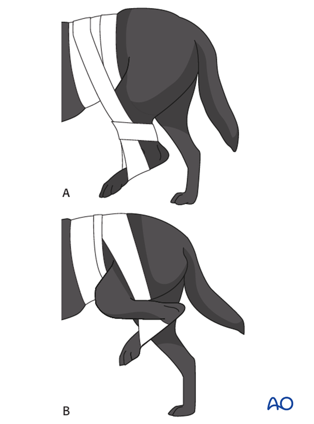
If after surgical treatment additional stability is required or the internal stabilization must be protected, a Robinson (A) or an Ehmer (B) sling may be applied.
This Robinson sling allows for a wide range of motion of the pelvic limb but prevents full weight bearing and full extension of the limb.
The Ehmer sling is used to slightly abduct and internally rotate the head of the femur. It restricts more mobility than the Robinson sling.
These two slings must be carefully monitored after application to avoid complications such as skin irritation, abrasions, swelling of the foot, and slippage of the sling.
Further information on the correct application of these slings can be found in the literature.
Implant removal
If there is no implant failure or infection, there is no need for implant removal.


