

Lag screw and K-wire
1. Principles
Fractures of the femoral neck are highly unstable because of:
- the long moment arm acting at the fracture plane (the entire femoral neck length), and
- the location of the fracture plane along the lines of maximum shear stress.
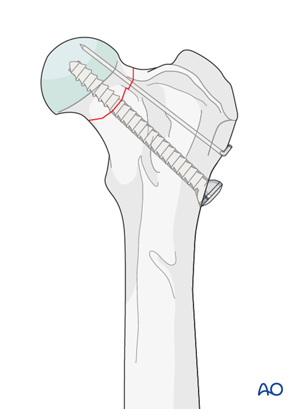
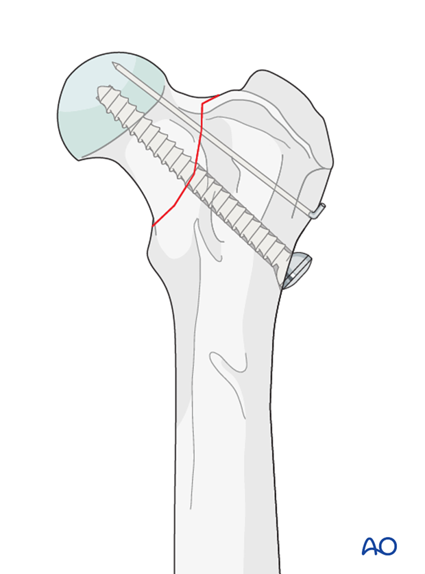
Compression of the fracture surface is needed to resist the high shear forces. Compression is achieved by using a lag screw, and stability is improved by adding an antirotational K-wire.
Although Salter-Harris type 1 fractures of the femoral head are generally treated with parallel K-wires only, physeal fractures in a more mature animal in which minimal growth potential remains can be stabilized using a lag screw and k-wires.
2. Patient positioning
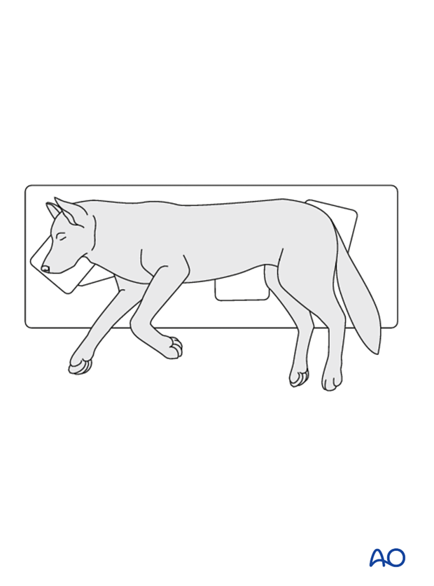
This procedure is performed with the patient in lateral recumbency.
3. Approach
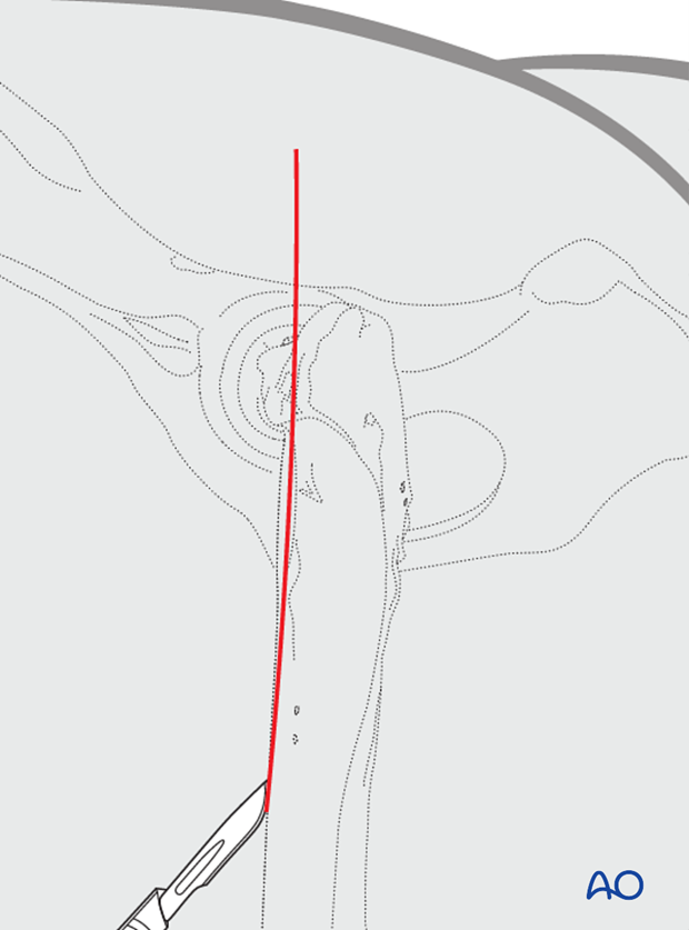
A craniolateral approach is performed.
4. Surgical technique
Implant selection
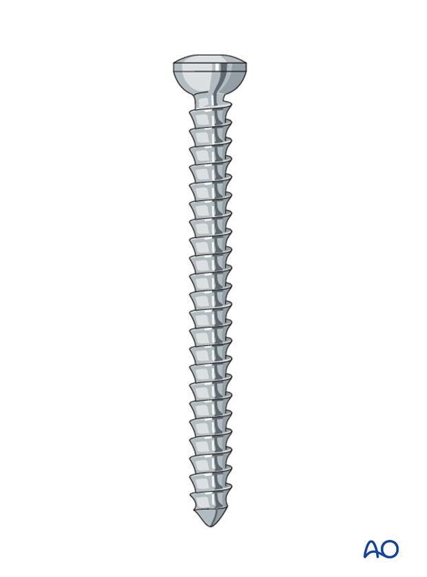
Although partially threaded screws or cannulated screws can be used, large cortical screws are often used in a lag fashion because of their large core diameter.
If a partially threaded screw is used, a screw with a large core diameter must be chosen. One must ensure that the threaded portion of the screw fully crosses the fracture plane to only engage the neck/head.
K-wire placement
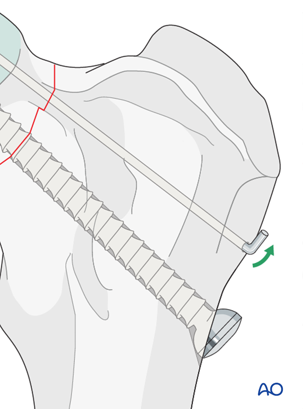
A K-wire is placed just proximal to the intended screw insertion site so it lays at the most proximal level of the fracture surface. Advancement of the wire stops when it reached the fracture surface.
The placement of the K-wire can be performed in a retrograde or normograde fashion.
Retrograde fashion ensures easier positioning of the pins within the femoral neck but requires a more aggressive exposure of the proximal femur to expose the fracture surface. A “C” guide is often used to accurately place the implants in a normograde fashion.
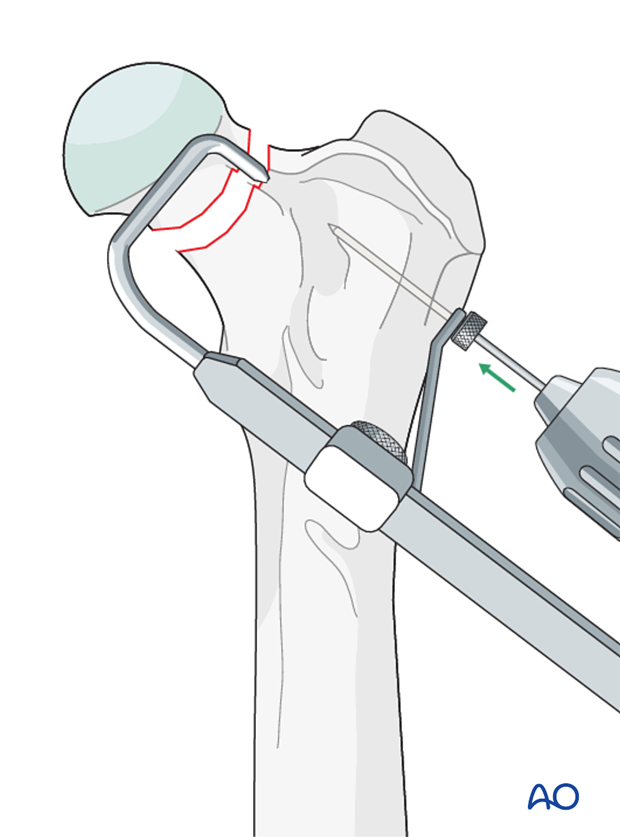
Reduction
The fracture is reduced and secured with pointed reduction forceps. The K-wire is driven into the femoral head without penetrating the articular surface.
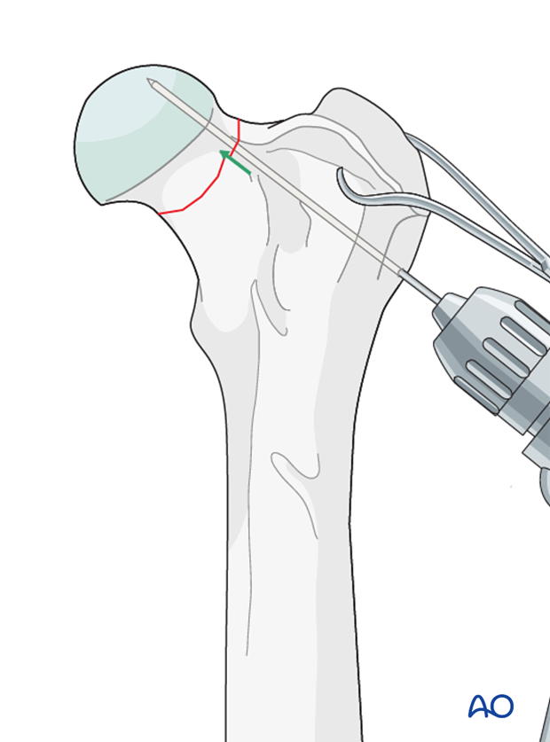
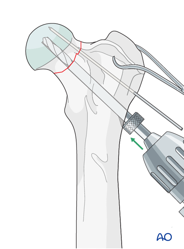
The glide hole for the screw is drilled into the femoral metaphysis below the level of the K-wire.
The insertion point for the screw on the femoral shaft is located just distal and slightly cranial to the third trochanter. The implants are oriented to follow the angle of inclination and anteversion of the femoral neck to ensure that the implants remain within the narrow femoral neck.
The drilling of the glide hole can be performed in a retrograde or normograde fashion.
Screw placement
The hole is measured and tapped if necessary. A screw just slightly shorter than the depth of the hole must be chosen to allow compression of the fracture before the tip of the screw contacts the cortex of the femoral head.
Although a partially threaded or a cannulated screw can sometimes be used, large cortical screws are preferred because of their large core diameter.
If a partially threaded screw is used, a screw with a large core diameter must be chosen and one must ensure that the threaded portion of the screw fully crosses the fracture plane and only engages the neck/head to achieve compression.
Read more about lag screw fixation.
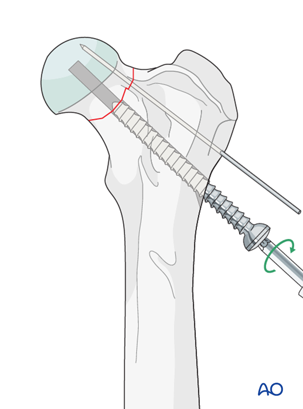
Pin is bent and cut short.
Validation of fixation
Postoperative orthogonal radiographs are taken to assess fixation.
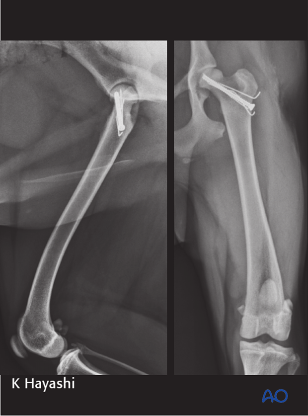
5. Case example Salter-Harris type I
1-year-old Australian cattle dog with a Salter-Harris type I fracture from falling.
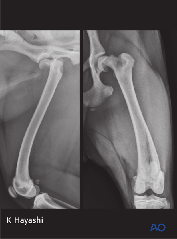
The fracture was repaired using a cannulated screw and three small pins.
Note: the distal femoral growth plate is closed and further growth from the proximal growth plate was minimal; hence a screw could be used.
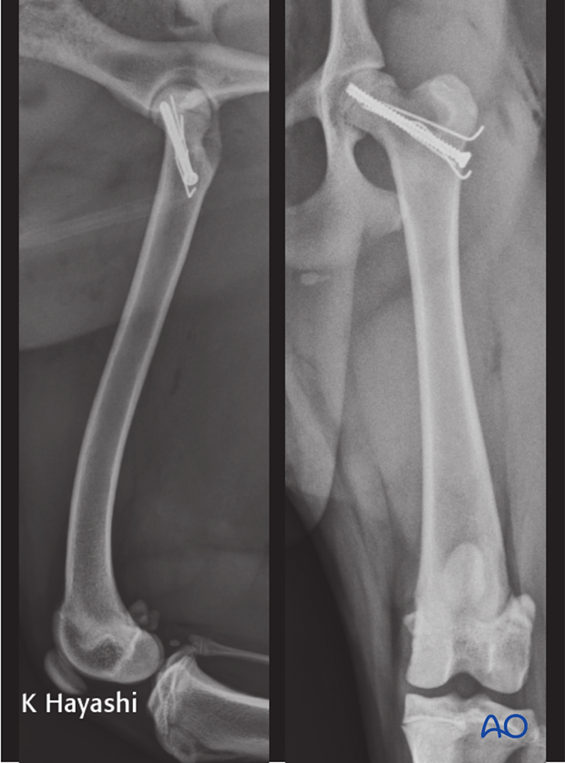
Postoperative radiographs at 2 months.
6. Aftercare
Activity restriction is indicated until evidence of bone union is detected on radiographic examinations.
Implants may cause discomfort of the adjacent soft tissue. If this occurs, implants are removed after radiographic evidence of bone healing is complete. In case of infection, implants must be removed after complete bone healing.
Phase 1: 1-3 day after surgery
Aim is to reduce the edema, inflammation, and pain.
Integrative medical therapies, anti-inflammatory and analgesic medications.
Phase 2: 4-10 days after surgery
Aim is to resolve the hematoma, edema and control pain, and prevent muscle contracture.
Anti-inflammatory and analgesic medications may still be needed. Rehabilitation and integrative medical therapies can be used.
Special attention should be given to patients less than 1 year of age with a femoral fracture. Rehabilitation is strongly recommended to help prevent quadriceps muscle contracture.
If the dog is not starting to use the limb within a few days after surgery, a careful evaluation is recommended.
10-14 days after surgery the sutures are removed.
Radiographic assessment is performed every 4-8 weeks until complete bone healing is confirmed.
Implant removal
If there is no implant failure or infection, there is no need for implant removal.


