Release notes - Dog radial and ulnar shaft
1. First edition
Publication of module (September 23, 2025)
The module on this anatomical area has been authored by Daniel Damur (Switzerland). Matthew J Allen (UK) acted as editor.
The dog radial and ulnar shaft module includes pages on:
- Fracture diagnosis
- Surgical approaches to the radial and ulnar shaft
- Patient positioning
- Complications
- Wide selection of fracture fixation techniques tailored to the specific fracture types
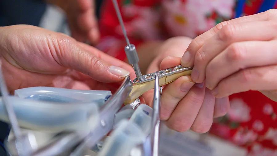
Around 300 original illustrations accompany the step-by-step descriptions of the various fixation techniques and approaches.
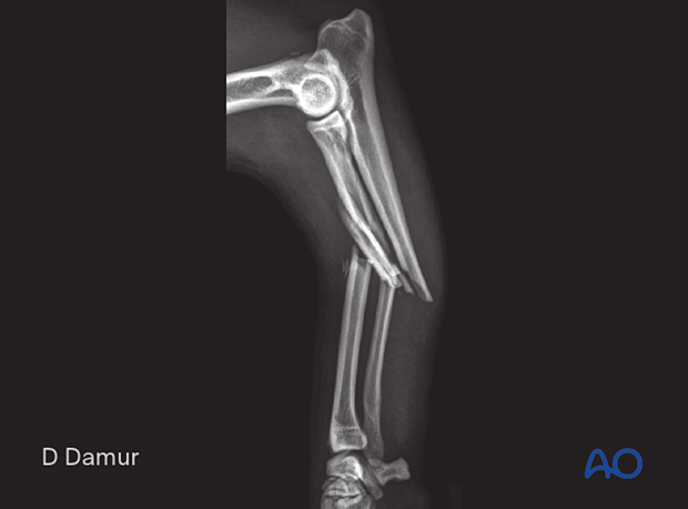
To help identify the fractures, x-rays of most fracture types have been included in the diagnosis section.
This module is exclusively for AO VET members.
Go to the module entry page.

Table of contents
The dog radial und ulnar shaft material includes a detailed description of the following fracture types and fixation methods.
22-A1 Incomplete fracture of the diaphyseal radius or fracture of one bone only:
- Nonoperative treatment
- Compression plate
- Linear external skeletal fixator
22-A2 Simple fracture of the distal zone of the diaphyseal radius:
- Nonoperative treatment
- Lag screws and neutralization plate
- Lag screw and neutralization plate on radius with IM pin or plate on ulna
- Compression T-plate
- Compression T-plate on radius with IM pin or plate on ulna
- Compression plate
- Compression plate on radius with IM pin or plate on ulna
- Circular or hybrid external skeletal fixator
- Linear external skeletal fixator
22-A3 Simple fracture of the proximal zone of the diaphyseal radius:
- Compression plate
- Compression plate on radius with IM pin or plate on ulna
- Lag screws and neutralization plate
- Lag screw and neutralization plate on radius with IM pin or plate on ulna
- Circular or hybrid external skeletal fixator
- Linear external skeletal fixator
- Bridging plate on radius with IM pin or plate on ulna
22-B1 Wedge fracture of the diaphyseal radius with simple ulnar fracture:
- Lag screws and neutralization plate
- Lag screw and neutralization plate on radius with IM pin or plate on ulna
- Bridging plate
- Bridging plate on radius with IM pin or plate on ulna
- Orthogonal bridging plates
- Linear external skeletal fixator
22-B2 Wedge fracture of the distal zone of the diaphyseal radius with multifragmentary ulnar fracture:
- Lag screws and neutralization plate
- Lag screw and neutralization plate on radius with IM pin or plate on ulna
- Bridging plate
- Bridging plate on radius with IM pin or plate on ulna
- Orthogonal bridging plates
- Linear external skeletal fixator
22-B3 Wedge fracture of the proximal zone of the diaphyseal radius with multifragmentary ulnar fracture:
- Lag screws and neutralization plate
- Lag screw and neutralization plate on radius with IM pin or plate on ulna
- Bridging plate
- Bridging plate on radius with IM pin or plate on ulna
- Orthogonal bridging plates
- Circular or hybrid external skeletal fixator
- Linear external skeletal fixator
22-C1 Complex fracture of the diaphyseal radius with simple or wedge ulnar fracture:
- Bridging plate
- Bridging plate on radius with IM pin or plate on ulna
- Orthogonal bridging plates
- Orthogonal bridging plates on radius with IM pin or plate on ulna
- Linear external skeletal fixator
- Circular or hybrid external skeletal fixator
22-C2 Segmental fracture of the diaphyseal radius with complex ulnar fracture:
- Bridging plate
- Bridging plate on radius with IM pin or plate on ulna
- Orthogonal bridging plates
- Orthogonal bridging plates on radius with IM pin or plate on ulna
- Linear external skeletal fixator
- Circular or hybrid external skeletal fixator
22-C3 Complex fracture of the diaphyseal radius with complex ulnar fracture:
- Bridging plate
- Bridging plate on radius with IM pin or plate on ulna
- Orthogonal bridging plates
- Orthogonal bridging plates on radius with IM pin or plate on ulna
- Linear external skeletal fixator
- Circular or hybrid external skeletal fixator
In addition, the material contains information about the following:
- Diagnosing each fracture type
- Indications for all treatments
- Patient positioning in dorsal recumbency, lateral recumbency, and sternal recumbency
- Craniomedial and MIO approaches to the radial shaft
- Caudolateral approach to the ulnar shaft
- Basic techniques for lag screw fixation, plate preparation, implant removal, and general considerations on external skeletal fixators


