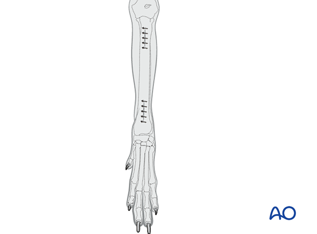
Minimally invasive osteosynthesis approach to the cat distal radius and ulna
1. Indications
The approach for minimally invasive osteosynthesis (MIO) is used for bridging plates.
2. Anatomy
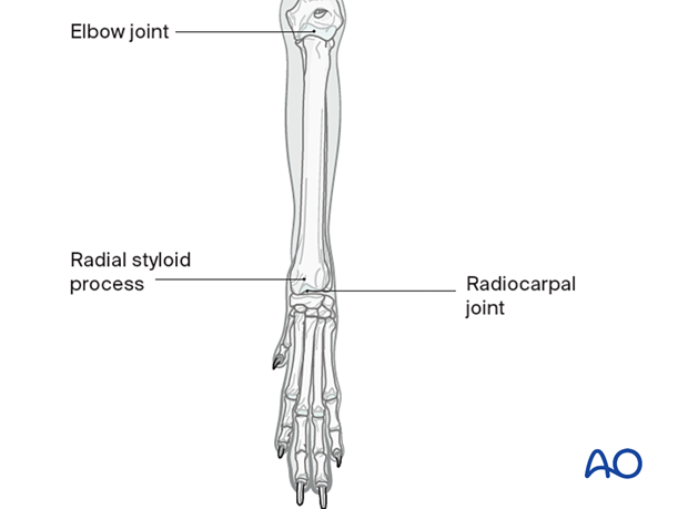
Key anatomical landmarks to be identified before the surgical approach include:
- Proximally, the elbow joint
- Distally, the radiocarpal joint and the radial styloid process
3. Patient positioning
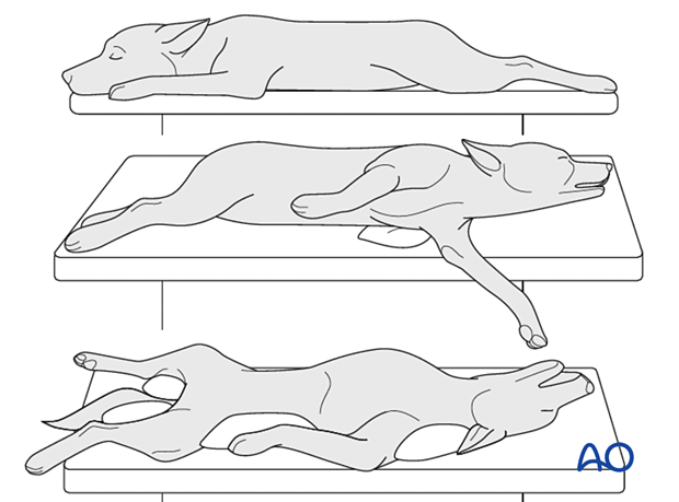
For this approach, the patient is positioned in one of the following positions:
4. Distal incision
Skin incision
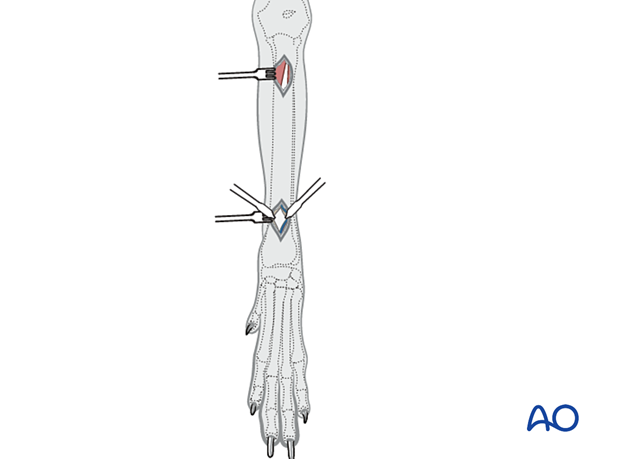
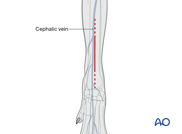
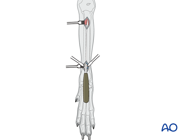
The distal incision extends proximally from just above the radiocarpal joint.
Exposure
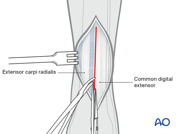
The deep antebrachial fascia is incised between the tendon of the extensor carpi radialis and the tendon of the common digital extensor.
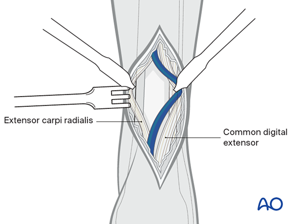
The tendon of the extensor carpi radialis is retracted medially and the tendon of the common digital extensor is retracted laterally to expose the dorsal surface of the distal radius.
5. Proximal incision
Skin incision
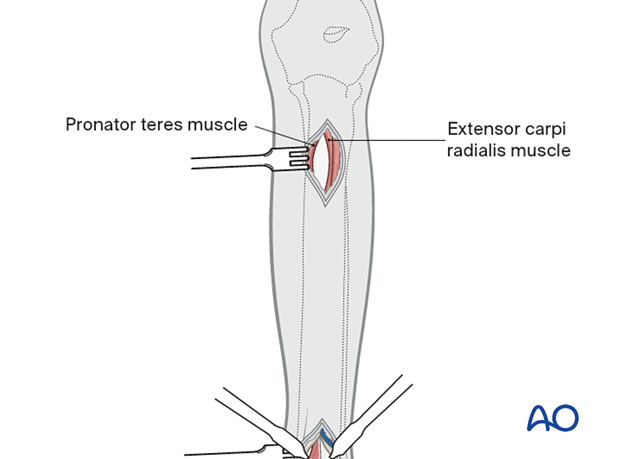
A cranial skin incision is made distal to the elbow joint line and over the body of the extensor carpi radialis muscle. The muscle bellies of the extensor carpi radialis muscle and the pronator teres muscle are separated by blunt dissection to expose the cranial shaft of the radius.
Exposure
The periosteal elevator is used to create an epiperiosteal tunnel connecting the proximal and distal incisions.
6. Closure
The subcutaneous tissue and skin are closed according to the surgeon's preference.