

Pancarpal arthrodesis with a plate
1. Indications
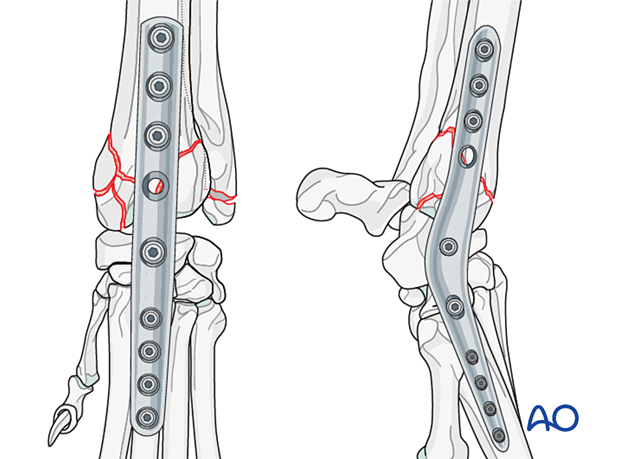
In cases of nonreconstructable articular fractures of the distal radius, salvage procedure with arthrodesis is required.
The metacarpal bones and the proximal radial fragment are realigned and stabilized in the correct spatial position, which provides rigid stability and anatomical alignment.
Plate application for pancarpal arthrodesis is preferred over external skeletal fixation because it is more acceptable to owners, less intensive in terms of management and associated with decreased risk of implant loosening.
2. Preparation and approach
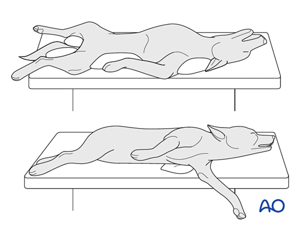
The patient can be in dorsal or lateral recumbency.
In lateral recumbency, the affected leg should be in contact with the table.
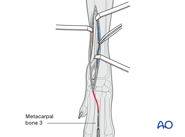
A craniomedial approach to the distal radius is extended on the dorsal side to the distal aspect of the third metacarpal bone.
3. Arthrodesis principles
The joint levels spanned by the implants are prepared by removal of the cartilage with a high-speed burr.
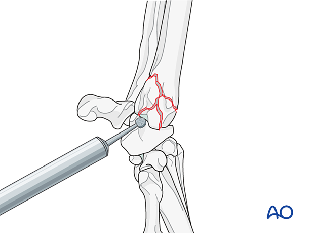
To promote bone fusion, autogenous or synthetic bone graft should be applied. Bone graft needs to be well distributed in each aspect of the antebrachiocarpal joint; this may require placement of bone graft after the radiocarpal screw is inserted.
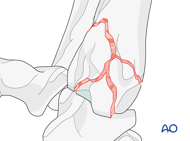
Implant selection
Commercially available, appropriately sized plates designed for medial or dorsal application are used for pancarpal arthrodesis.
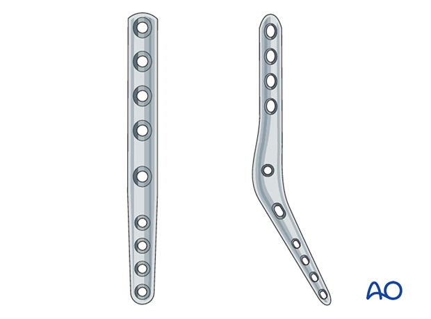
Dorsal plating
An appropriately sized plate is contoured to provide 5–10 degrees of extension of the radiocarpal joint.
Arthrodesis plates are generally hybrid plates allowing for insertion of smaller screws in the metacarpal bones and larger screws in the radius.
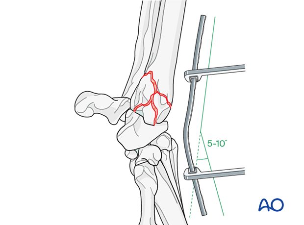
The plate is centered over the dorsal surface of the third metacarpal bone and the first screw is inserted in the radial carpal bone.

The next screw is inserted in the most distal aspect of the third metacarpal bone.
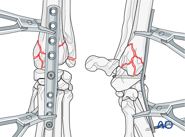
The third screw is inserted in the most proximal aspect of the third metacarpal bone.
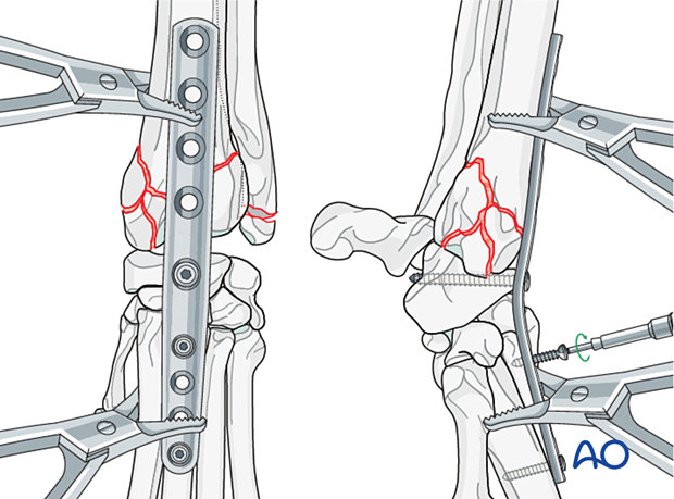
The fourth screw is placed in the distal radius, close to the fracture line.
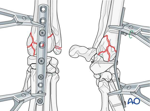
The remaining holes are filled with screws.
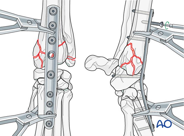
Medial plating
An appropriately sized plate is contoured to provide 5–10 degrees of extension of the radiocarpal joint. In order to achieve this, a plate that can be contoured in three planes is required. Alternatively, dedicated precontoured anatomically designed plates are available.
Arthrodesis plates are generally hybrid plates allowing for insertion of smaller screws in the metacarpal bones and larger screws in the radius.
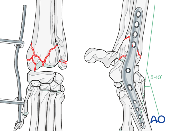
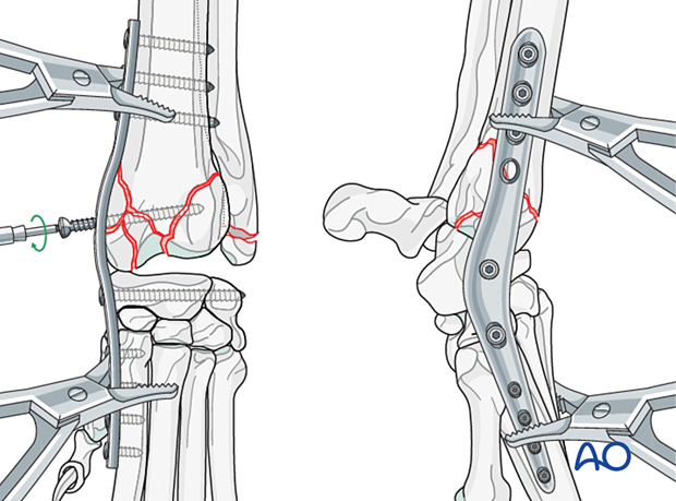
The plate is positioned over the medial surface of the distal radius, spanning the carpus and reaching the medial surface of the second metacarpal bone. To make plate contouring easier, the styloid process of the radius can be removed with an oscillating saw or a high-speed burr.
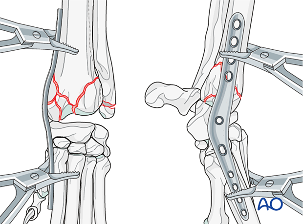
The first screw is inserted in the radial carpal bone.
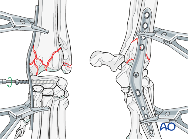
The next screw is inserted in the most distal aspect of the metacarpal bone.
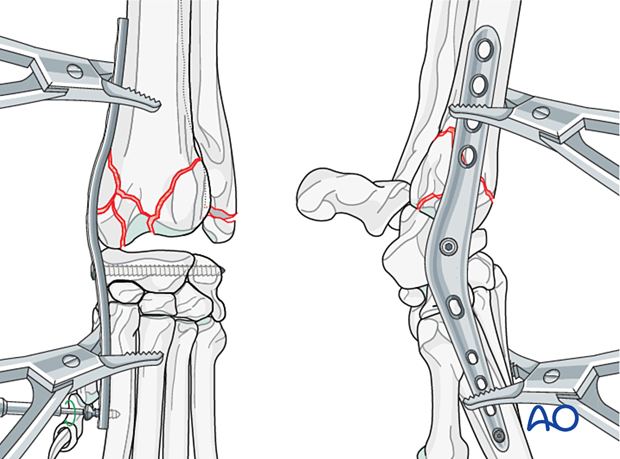
The third screw is inserted in the most proximal aspect of the metacarpal bone.
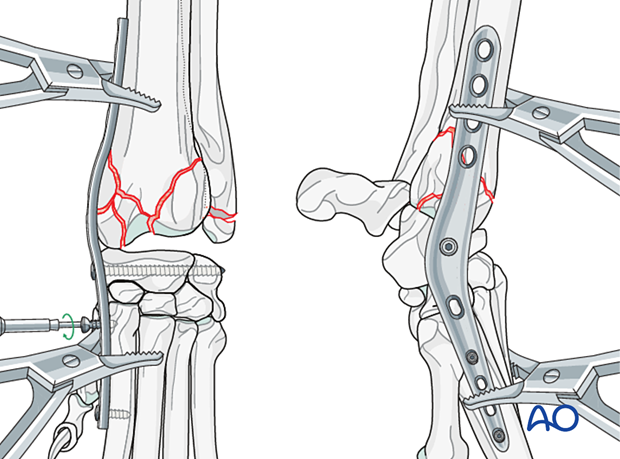
The fourth screw is placed in the distal radius close to the fracture line.
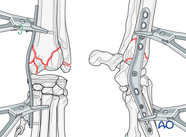
The remaining holes are filled with screws.
4. Aftercare
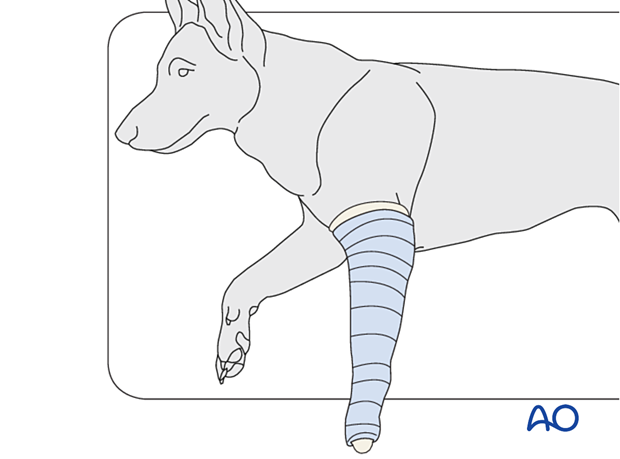
A Robert Jones or modified Robert Jones bandage can be applied for 1–3 days to decrease the edema and protect the surgical incision.
Phase 1: 1–3 days after surgery
The aim is to reduce edema, inflammation, and pain.
Integrative medical therapies, anti-inflammatory medications, and analgesics are recommended.
10–20 minutes of ice therapy is recommended every 8 hours in most cases.
The patient can immediately bear weight, but with strict control of activities.
Phase 2: 4–10 days after surgery
The aim is to resolve hematoma and edema, control pain, and prevent muscle contracture. Anti-inflammatory and analgesic medications may still be needed.
Rehabilitation and integrative medical therapies can be used.
A careful evaluation is recommended if the patient is not starting to use the limb within a few days after surgery.
Phase 3: > 10 days after surgery
The sutures are removed 10–14 days after surgery.
Immature patients should be radiographed every 4 weeks.
A radiographic assessment is performed every 4–6 weeks until bone healing is confirmed, more frequent assessment may be necessary for skeletally immature patients.


