

K-wire with or without tension band
1. Indications
The goal of 23-A1 fracture treatment is anatomical reduction.
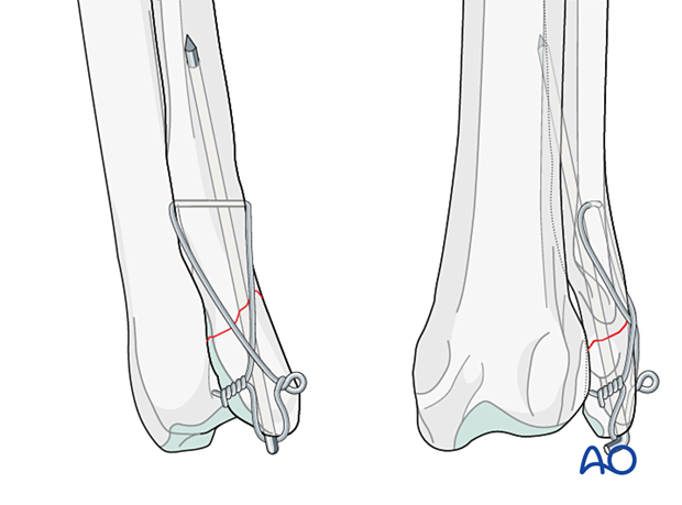
This is achieved by inserting a K-wire from the distal fragment across the fracture line and placing a tension band wire in a figure-of-eight pattern over the proximal and distal fragments.
K-wire fixation with or without tension band wire is indicated for isolated distal ulnar fractures (also with small fragments) in small to medium breed dogs and cats.
2. Preparation and approach
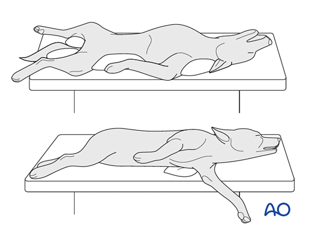
The patient can be in dorsal or lateral recumbency.
In lateral recumbency, the nonaffected leg should be in contact with the table.
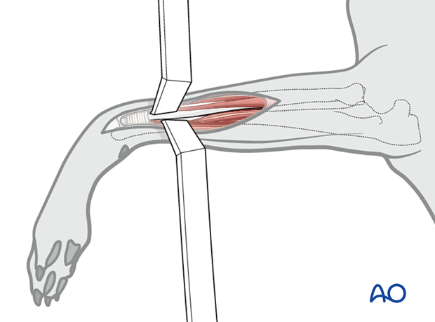
A lateral approach to the distal ulna is performed.
3. Reduction
The goal is to achieve anatomical reduction.
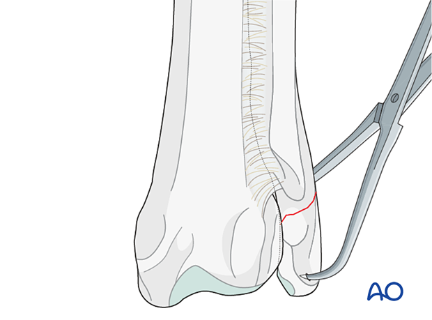
The proximal fragment is connected to the radius through interosseous ligament and is stable, not requiring manipulation.
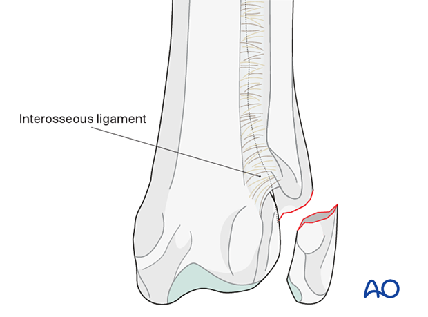
The distal fragment is manipulated with a pointed reduction forceps to achieve reduction.
Option
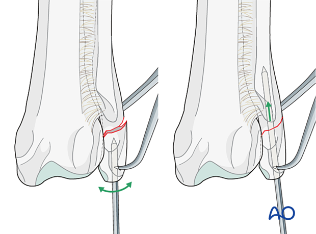
Alternatively, the K-wire can be inserted in the distal fragment first and then used as a joystick to achieve reduction.
4. Fixation
Implant selection
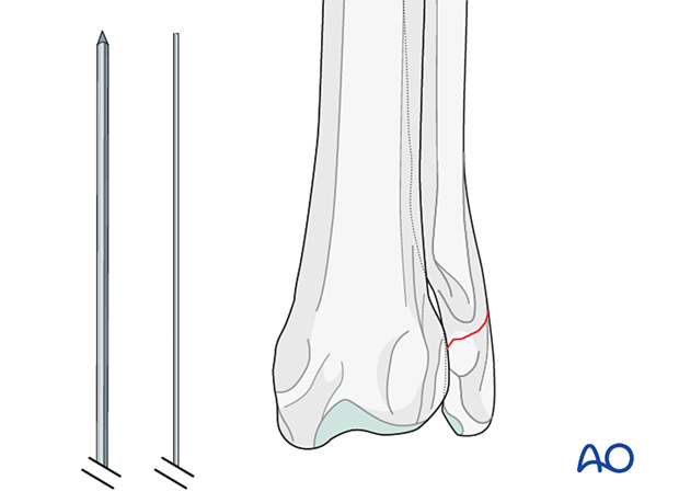
Appropriately sized K-wire(s) and tension band wire are selected.
In most cases, it is not possible to place two K-wires and a single K-wire can provide enough stability.
K-wire insertion
A single K-wire is inserted normograde from the distal fragment across the fracture line into the medullary canal of the proximal fragment to achieve reduction.
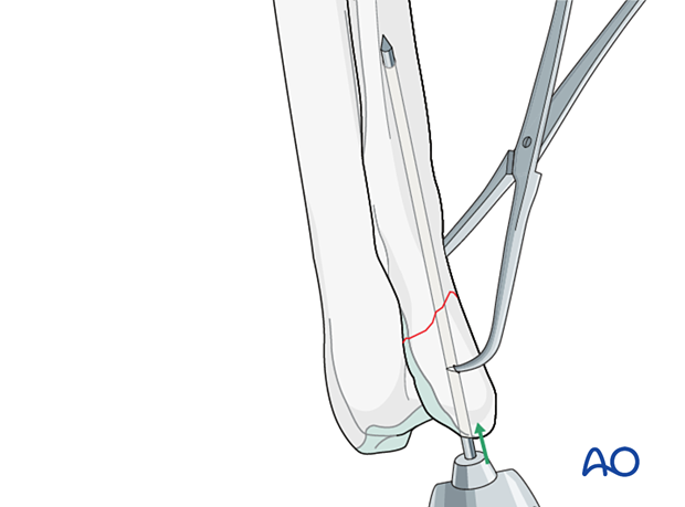
In bigger patients, a second K-wire can be inserted in parallel direction.
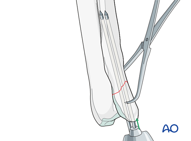
Tension band
A small hole is drilled from cranial to caudal in the proximal fragment. The diameter of the drill hole should be optimized to the size of the tension band wire. The tension band wire diameter is selected to approximate the size of the K-wires.
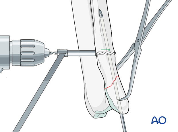
The tension band wire is inserted through the hole, and the ends are crossed on the lateral surface of the ulna.
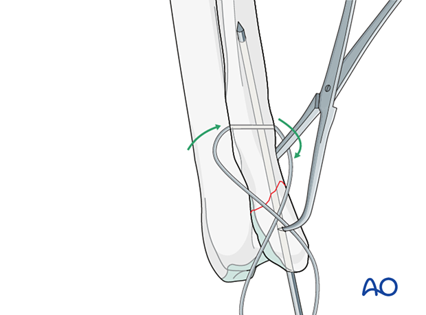
The wire is passed around the ends of the K-wires and back to the other end of the wire on the starting side of the bone. This creates a figure-of-eight pattern.
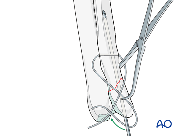
Twist knots are commonly used for tension band wires, although loop-style knots can also be used.
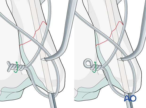
To effectively tighten a tension band wire with one knot, the tightening process must also draw in the slack in the arm opposite the one with the knot. Because the wire makes several tight bends, both through the hole in the bone and around the K-wires, this may not happen with larger diameter wire.
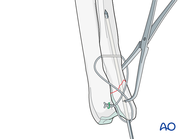
A double knot technique is preferred to address this deficiency of the single knot technique.
A twist knot is tied in both arms of the figure-of-eight. When placing the wire, a loop is formed in the wire so that it is positioned between the hole and the K-wires in the first arm of the figure-of-eight.
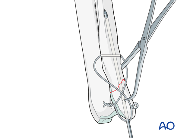
The tension band wire is tightened by twisting the two knots.
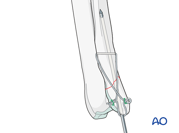
Once the slack has been removed, the K-wires are bent over so that they lie flat to the bone. The bent K-wires are directed away from the pull of the wire by bending them away from the bone, cutting them with 2–3 mm of the bent arm, and rotating them to direct the arm away from the wire. The end will usually embed in the soft tissues around the fragment.
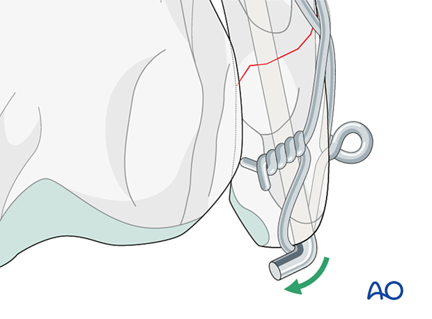
Once the final position of the K-wires is set, tightening of the figure-of-eight is completed.
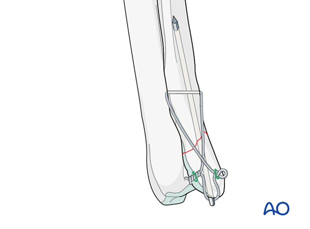
In some cases, in smaller patients, K-wire without tension band wire can be an option and can be combined with a splinted bandage.
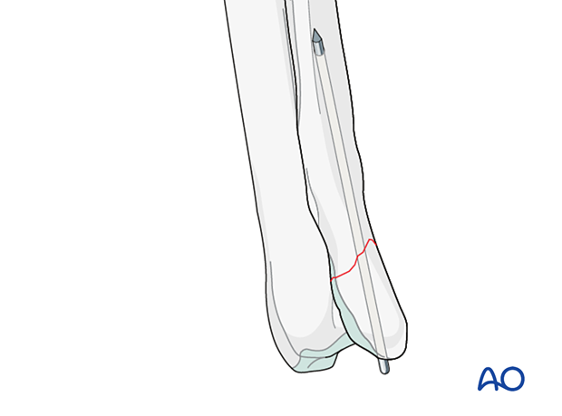
5. Aftercare
Phase 1: 1–3 days after surgery
The aim is to reduce edema, inflammation, and pain.
Following appropriate internal fixation, there should be no absolute need for external coaptation for a 23-A1 fracture. If bandaging is used to decrease edema and protect the surgical wound, it should be removed after 24–48 hours to reduce the risk of complications.
Integrative medical therapies, anti-inflammatory medications, and analgesics are recommended.
10–20 minutes of ice therapy is recommended every 8 hours in most cases.
The patient can immediately bear weight, but with strict control of activities.
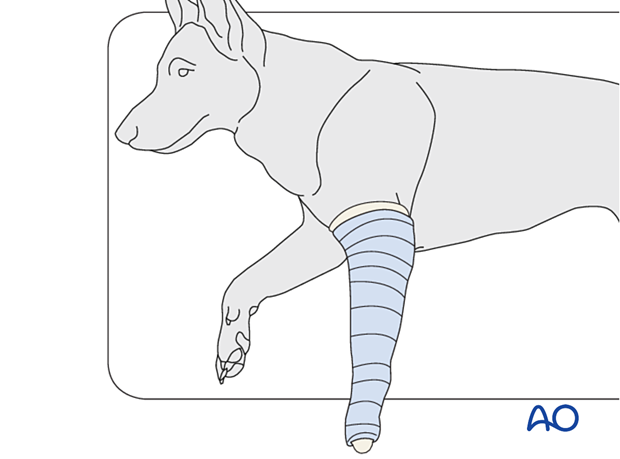
Phase 2: 4–10 days after surgery
The aim is to resolve hematoma and edema, control pain, and prevent muscle contracture. Anti-inflammatory and analgesic medications may still be needed.
Rehabilitation and integrative medical therapies can be used.
A careful evaluation is recommended if the patient is not starting to use the limb within a few days after surgery.
Phase 3: > 10 days after surgery
The sutures are removed 10–14 days after surgery.
Immature patients should be radiographed every 4 weeks.
A radiographic assessment is performed every 4–6 weeks until bone healing is confirmed, more frequent assessment may be necessary for skeletally immature patients.


