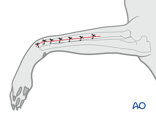
Lateral ulnar approach to the cat distal radius
1. Indications
An open approach to the lateral surface of the ulna is used for procedures requiring exposure and reduction of the distal ulna.
This approach can also be used for an “open-but-do-not-touch” approach, allowing fracture visualization and indirect reduction without removing the fracture hematoma and manipulating the fragments.
2. Anatomy
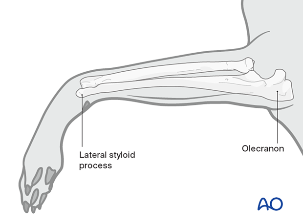
Key anatomical landmarks to be identified before the surgical approach include:
- Proximal olecranon
- Lateral styloid process
3. Patient positioning
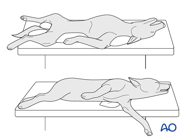
For this approach, the patient is positioned in one of the following positions:
- Lateral recumbency with affected leg up
- Dorsal recumbency
4. Skin incision
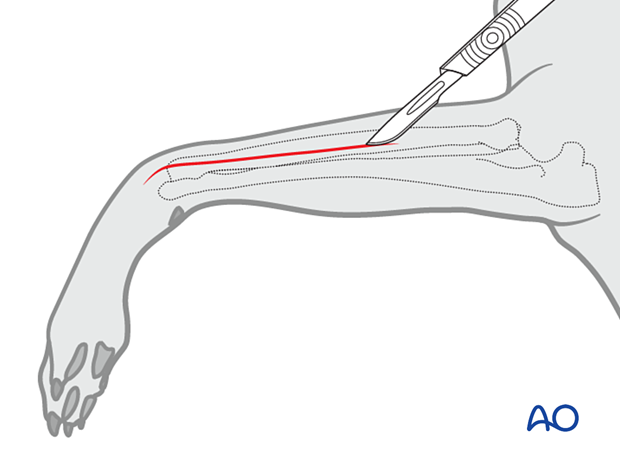
A lateral skin incision is made proximal to distal to expose the fracture area. The incision extends along a line connecting the lateral humeral condyle to the ulnar styloid process.
The final length of the incision should be dictated by the procedure that is being performed.
5. Development
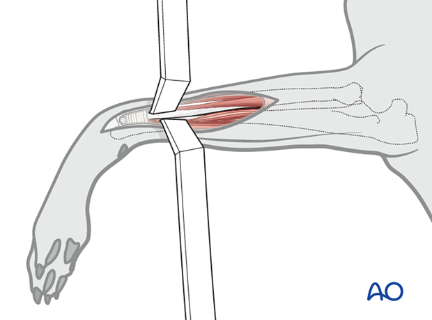
The subcutaneous tissue is dissected along the same line as the skin.
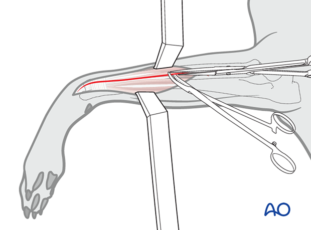
The distal ulna is exposed by retraction of the tendons of the common digital extensor and lateral digital extensor muscles.
6. Wound closure
The subcutaneous tissue and skin are closed according to the surgeon's preference.