Anterior lumbar interbody fusion (ALIF)
1. Introduction
ALIF is one of the best less invasive surgeries for anterior support in the distal part of the lumbar spine.
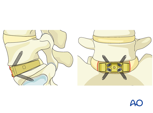
Approach
This procedure is the best approach for level L5–S1.
This procedure can be used at the levels above (L4–L5 and L3–L4) but is technically more difficult due to the anatomical configuration with its close relationship to the great vessels (abdominal aorta and vena cava).
An approach surgeon is advised if the surgeon is unfamiliar with this procedure.
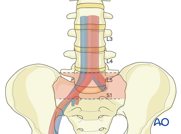
Mandatory MRI evaluation
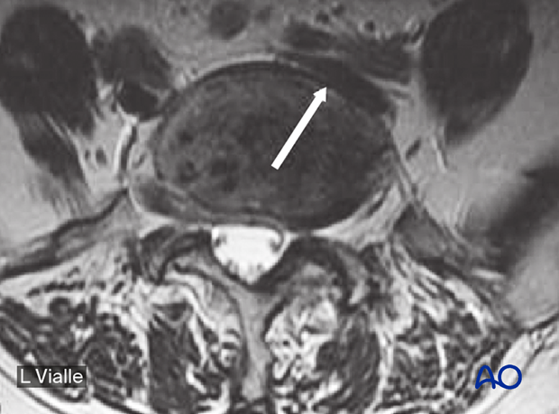
An MRI evaluation of the vessels is mandatory.
The aorta bifurcation and the position of the iliac veins must be identified before the procedure.
In this image, the arrow shows the left iliac vein.
2. Preparation
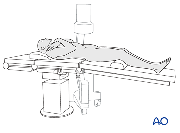
The patient is positioned in a supine position with arms folded across the chest. A slight opening of the table in lordosis is recommended.
3. Fluoroscopic identification of the target level before draping
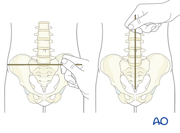
The correct operative level is determined using fluoroscopy.
The C-arm should be perfectly adjusted to perform real AP and LL views (the endplate of L5 should be exactly identified and lie on the same line).
The spinous process and end plates must be aligned according to the patient’s lordosis.
It is helpful to use a localizer on the skin to identify the level and the incision position.
4. Approach
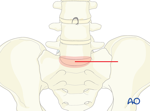
This procedure is performed through the retroperitoneal approach.
5. Disc removal
Identify the lateral borders of the L5–S1 disc.
Incise the anterior annulus midline in a vertical fashion and develop a flap on either side, whenever possible.
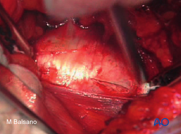
Proceed to remove the disc down to the subcentral bone. Care must be taken not to enter the epidural space.
The use of laminar spreaders will facilitate disc removal.
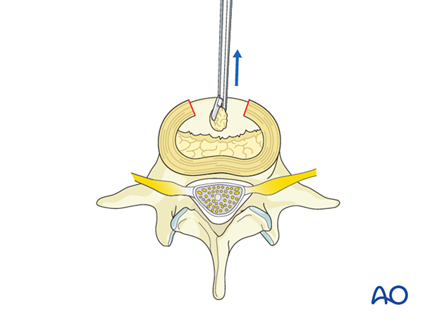
Meticulously remove the entire nucleus pulposus and all the cartilage from the endplates. Pieces of cartilage may inhibit fusion if present either on the endplates or in the bony material used for the fusion.
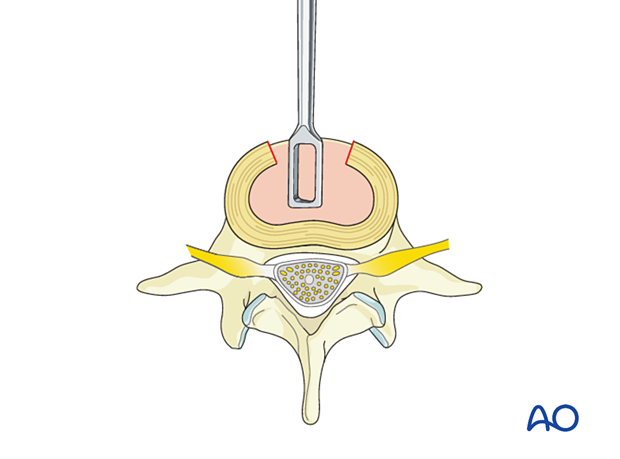
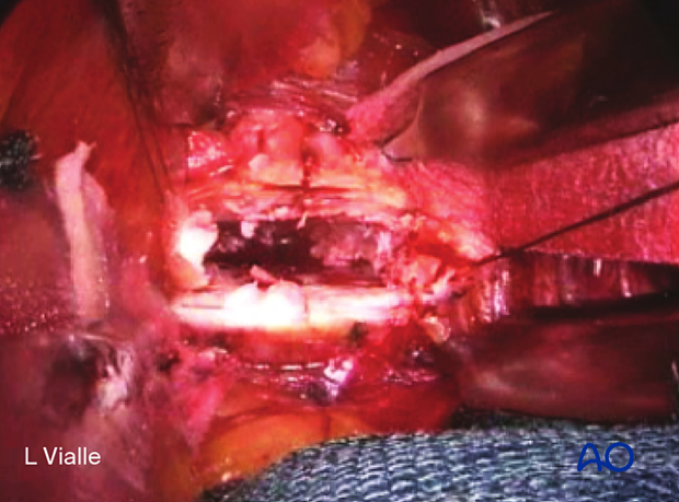
The disc nucleus is totally removed.
The image shows flaps with a retainer suture on each side.
6. Cage selection
The estimated cage size can be determined using measurements from the patient's image. The ideal cage size should cover approximately 90% of the endplate.
The cage should be firmly seated in the disc space.
Test cages are used to determine the correct cage size.
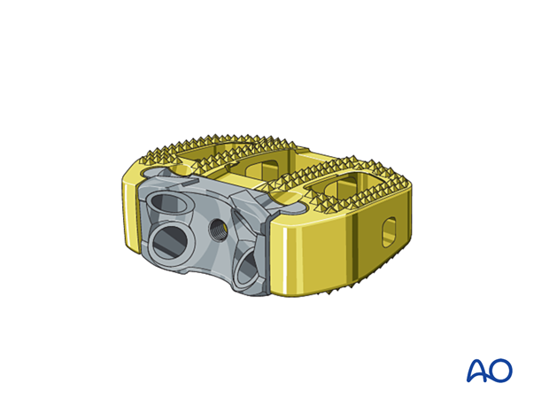
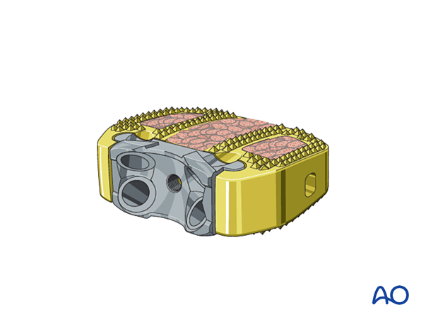
The cage must be filled with bone or bone substitute.
7. Cage insertion
By breaking the operative table, a mild increase in the lordotic configuration is produced. The cage is then inserted, and the cage inserter is struck properly until the desired position is reached. This procedure is controlled by the C-arm in AP and lateral positions.
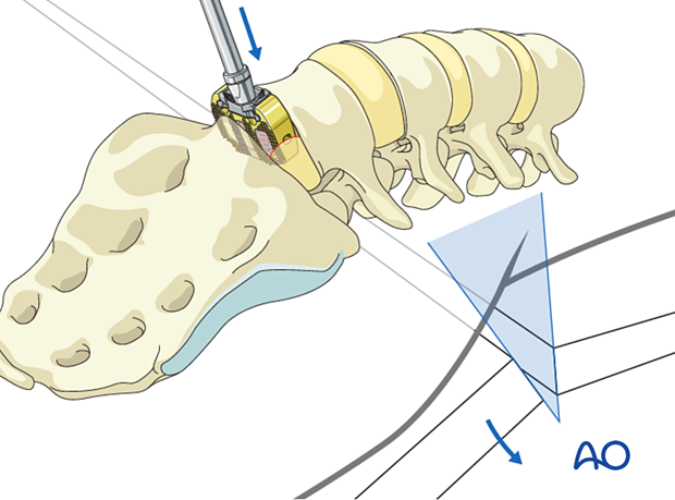
After the final positioning of the cage is achieved, the lordosis is reduced by returning the table to the initial position.
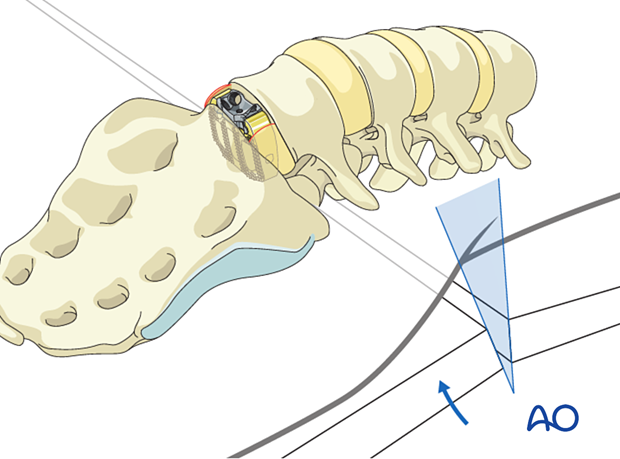
A set of anchors or screws can be attached, ensuring sound fixation and stabilization.

8. Additional instrumentation
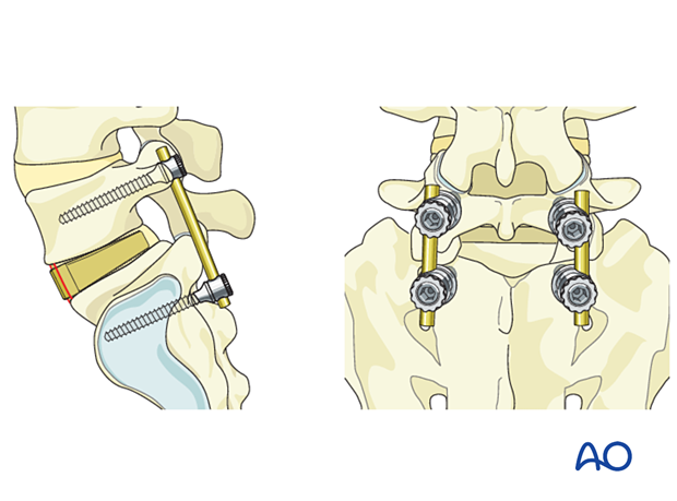
Additional fixation is recommended if stand-alone cages are used (if the stabilizers mentioned previously are not used).
Open or percutaneous screws can be used according to preoperative planning.
9. Closure
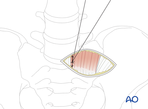
After careful visualization of all the anatomical structures that have been passed through during the approach, the fascia can be closed. The remainder of the wound should be sutured in a standard fashion, usually without using drainage.
10. Aftercare
Patients can usually stand on the first day after surgery and can be discharged on the second day after surgery.
For the first month after surgery, it is recommended that a brace is used to reduce the movements of the lumbar spine.
Postoperative physiotherapy is not essential but can be useful in some selected cases.


