Prone position for approaches to C0–C7
1. Patient positioning
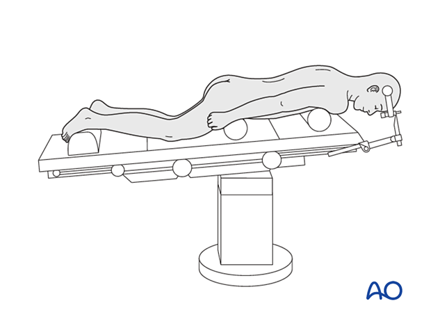
The patient is positioned prone on a radiolucent table with two horizontally placed padded bolsters (one at the level of sternum and another one at the level of anterior iliac spine) or a Jackson table frame.
- The head is securely fixed in a Mayfield three-pin head holder. This avoids pressure on the eyes.
- The head is slightly flexed to increase the foraminal area.
- The chin should be free and not touching the table.
- The abdomen should hang free to avoid increased intraabdominal pressure to prevent excessive bleeding.
- The arms are positioned downwards at the sides, and the elbows and wrists are padded to prevent peripheral neuropathies. The arms are secured with tape or a sheet.
- Gentle traction on the shoulders can help with visualization, especially at the lower cervical levels.
- A small bump or blanket roll should be placed under the legs to flex the knees slightly.
- Adequate padding must be provided to the knees and dorsum of the feet to avoid pressure sores.
- The table is placed in a reversed Trendelenburg position.
- The patient should be adequately secured to the table.
The Mayfield head clamp should be applied while the patient is supine. When using a Mayfield clamp, select the pin entry points so that the clamp can be rotated freely over the nose once the patient is put into the prone position.
Make sure that there are adequate personnel to receive and turn the patient from a supine to a prone position on the operating table as needed.

2. Anesthesia
General anesthesia with endotracheal intubation is performed.

3. Preoperative antibiotics
Antibiotics should be administered prior to incision and at two-hour intervals during the procedure.
A cephalosporin antibiotic with good Gram-positive coverage is generally recommended.
Patients with penicillin allergies should receive vancomycin or clindamycin.
4. Spinal cord monitoring
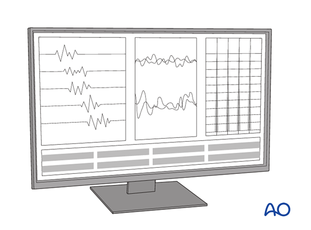
Spinal cord monitoring can be used with SSEP, MEP, and free-running EMG. In select cases, baseline monitoring should be considered before flipping the patient, and performed again after final positioning to confirm stable spinal cord monitoring.

5. Fluoroscopy
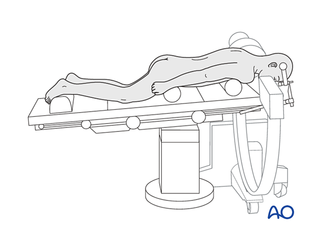
The incision can be planned based on AP and lateral x-rays.
An intraoperative CT scan can be used together with spinal navigation.

OR Setup
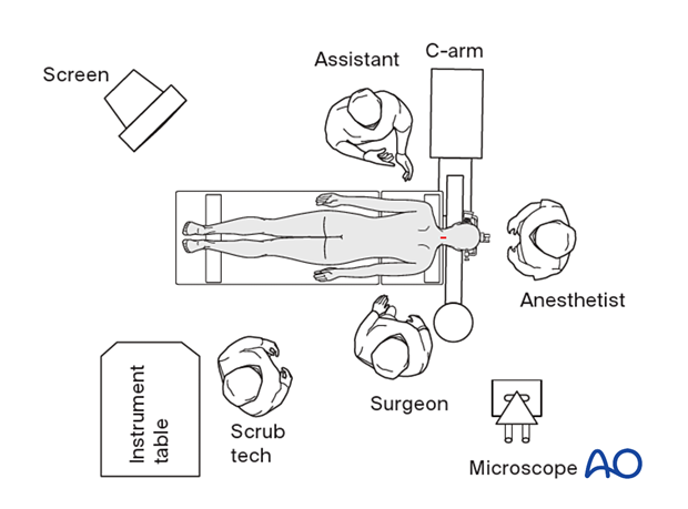
The description above is the setup for MIS.
If a microscope is used, it should be placed on the surgeon’s side and opposite the image intensifier portion of the C-arm fluoroscope.


