Posterior fusion of L4-S1 (with or without pelvic fixation or ala)
1. Introduction
Basic principles
Managing high grade spondylolisthesis requires good knowledge of lumbosacral anatomy.
The goals of the surgical management are:
- Solid fusion across the deformity
- Neural decompression
- Ensure sagittal balance
- Prevent further slippage
- Reduce pain
- Maintain neurological integrity
Identification of anatomical landmarks
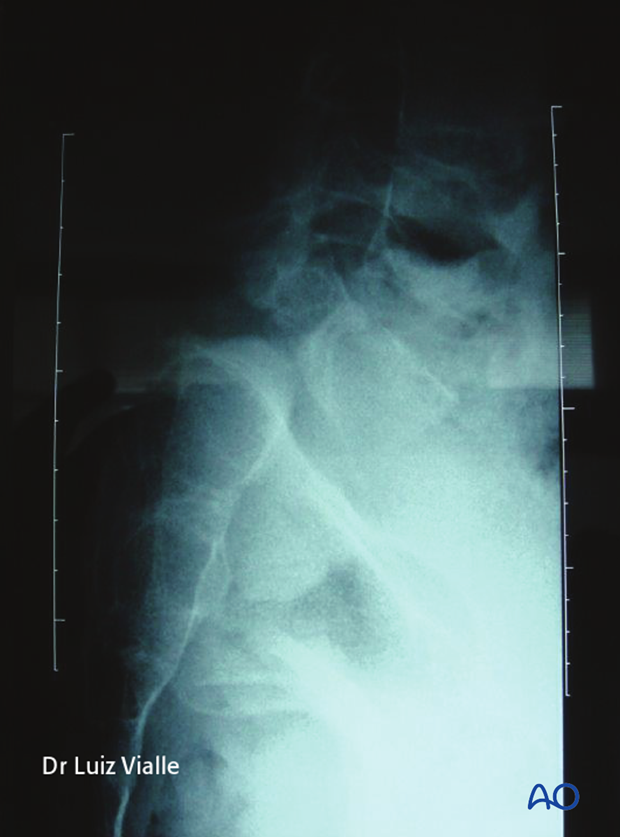
Due to the distorted anatomy care must be taken to confirm correct fusion levels. Typically the L5 pedicle is extremely anterior, hidden beneath the sacral alar.
In high grades it recommended to span the fusion from L4 to S1 or pelvis.
Intraoperative fluoroscopy or spinal navigation is used to facilitate identification of correct levels.
2. Prepartation and approach
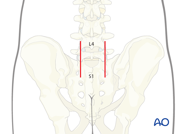
This procedure is performed through the Wiltze approach with the patient placed prone.
3. Pedicle screw insertion
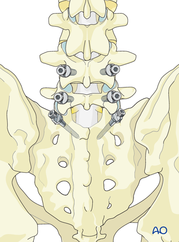
Pedicle screws are inserted in L4, L5, S1, and/or in the iliac crest in a standard fashion, ensuring that the proximal facet of L4 is not breached by the pedicle screw.
We will here show the fixation of L4-S1.
4. Spine decompression
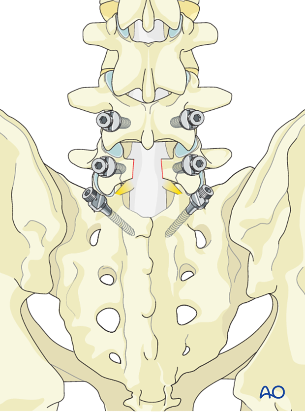
A wide posterior spinal decompression is performed by carefully removing the L5 mobile lamina and the ligamentum flavum using sequential Kerrison rongeurs. The dural sac and the L5 and S1 nerve roots are identified.
5. Reduction
Facet removal
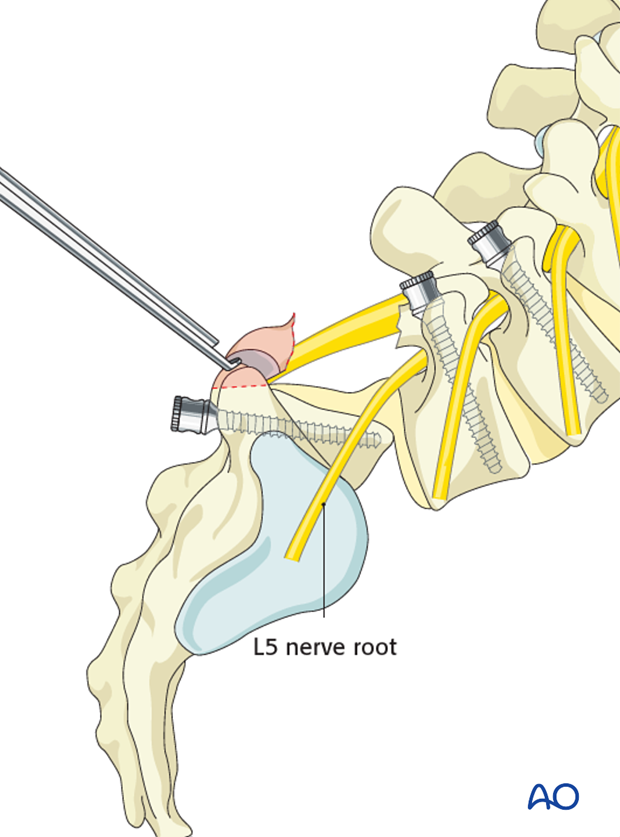
Due to the anterior listhesis the course and location of L5 nerve root places it at risk. Great care is therefore taken to identify and protect it during facet and disk removal.
The inferior facet of L5 and part of the S1 superior facet is removed bilaterally to give access to the L5-S1 disk space.
Care is taken not to injure the L5 nerve.
Disk removal
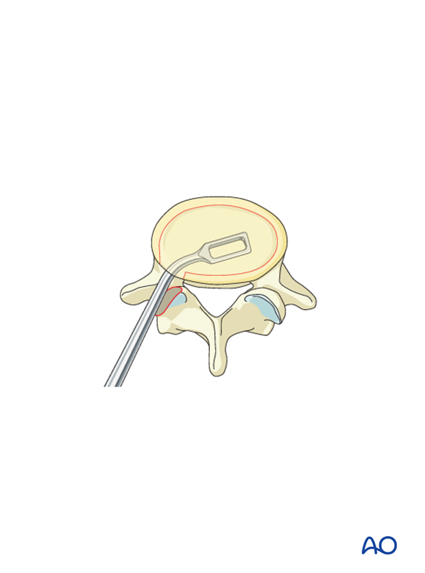
The disc is removed by incising the anulus fibrosis and using a series of rongeurs and curettes forward to the anterior anulus. Care is taken not to disrupt the anterior anulus.
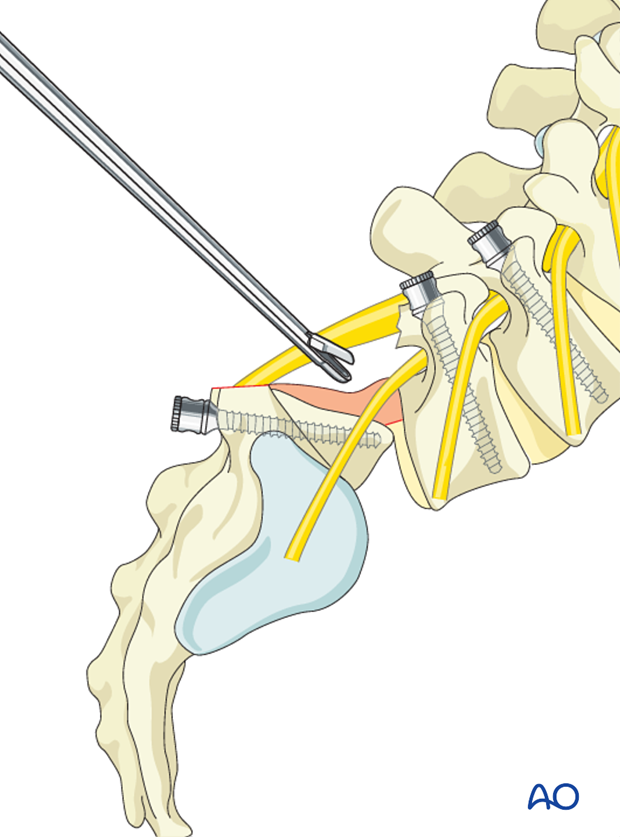
The endplates are cleared for all traces of cartilaginous material to ensure fusion.
If the sacral endplate is domed, the dome is flattened using osteotomes or curettes.
Mobilization of disk space
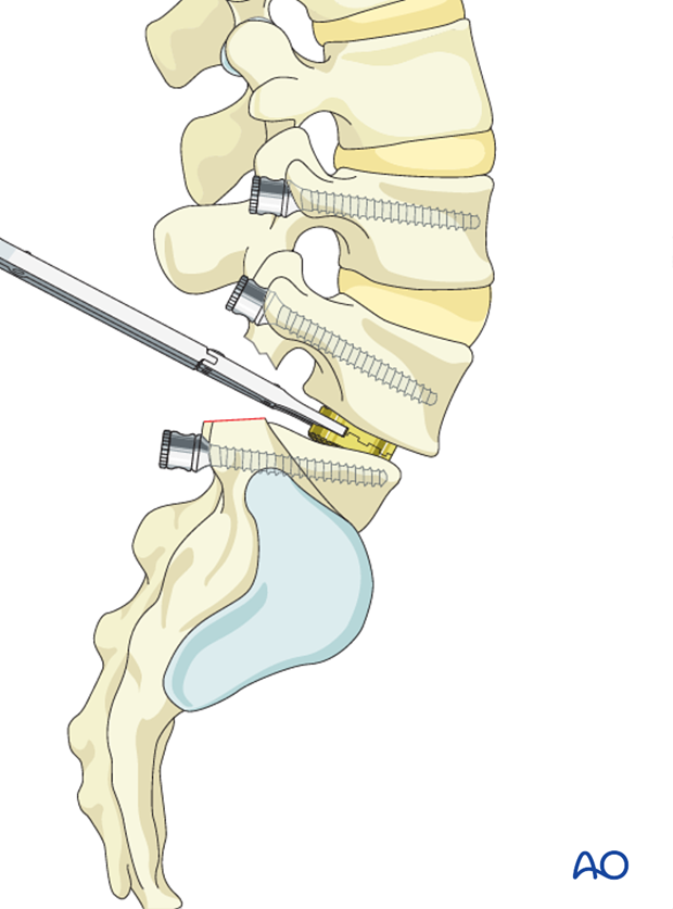
Interbody trials / spacers are inserted bilaterally in incremental fashion to loosen the annulus and anterior longitudinal ligament to achieve indirect foramina decompression by increasing the inter vertebral hight.
The focal lumbosacral kyphosis must be corrected during this maneuver.
The Listhesis can also be corrected to improve anterior vertebral apposition however care must be taken as anatomical reduction has been associated with L5 nerve root injury.
During this maneuver the L5 nerve roots EMG's must be monitored to avoid postoperative foot drop.
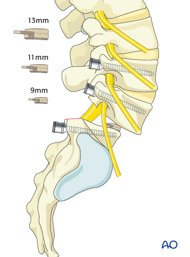
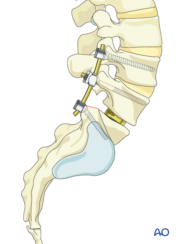
Once the satisfactory reduction is achieved, the spacer is replaced by the corresponding cage.
Care must be taken to ensure that the cage is resting between both endplates. A structural graft can also be used instead of a cage.
The graft or cage should be placed as anterior as possible in order to achieve more lordosis.
High grade listhesis are at higher risk of graft extrusion.
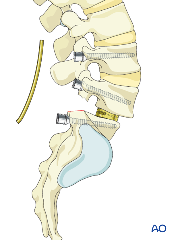
Rods are contoured to accommodate the pedicle screws and the iliac screws. The rods must respect the achieved regional lordosis.
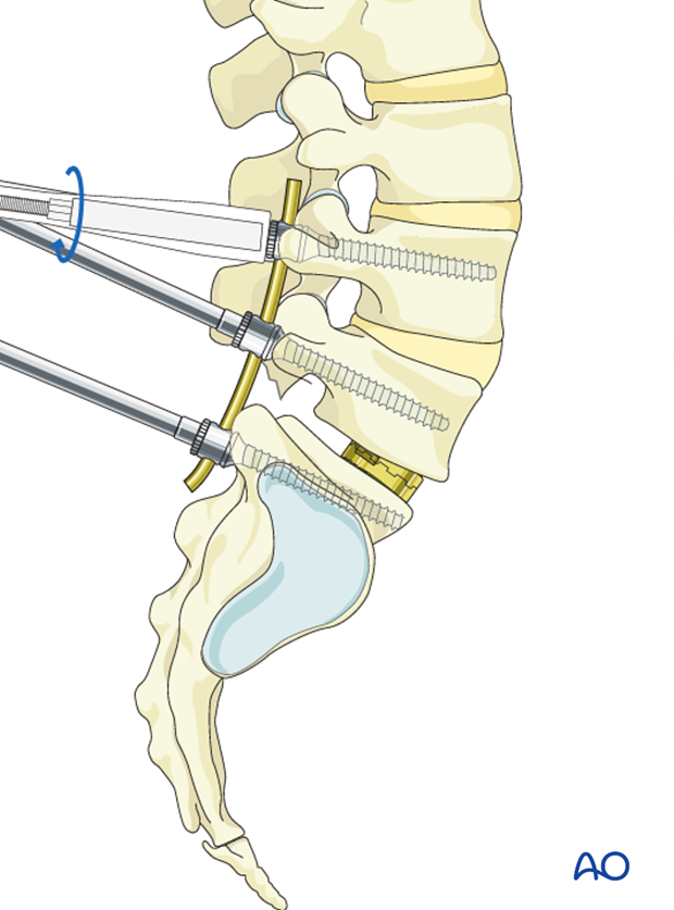
The achieved correction, listhesis and kyphosis are stabilized by securing the rod to the screws.
Optional: reduction with Schanz pins
Special 6 mm double-threaded Schanz screws are inserted in the L5 pedicle on each side. The Schanz screws have a 3 cm long threaded part in their shaft.
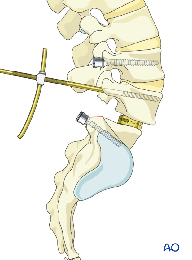
Two rods about 8 cm long are contoured to the desired lumbosacral lordosis. The rods are mounted to the Schanz pins in L5 with a rotule and then the rods are pushed into the screws of L4 and S1 where they are fixed.
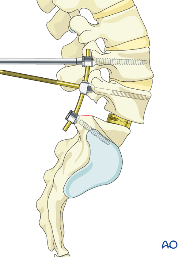
Two threaded sleeves are sleeved over the Schanz screws. By tightening the sleeve along the threaded part of the Schanz screws, the vertebra of L5 is gradually reduced towards the rod.
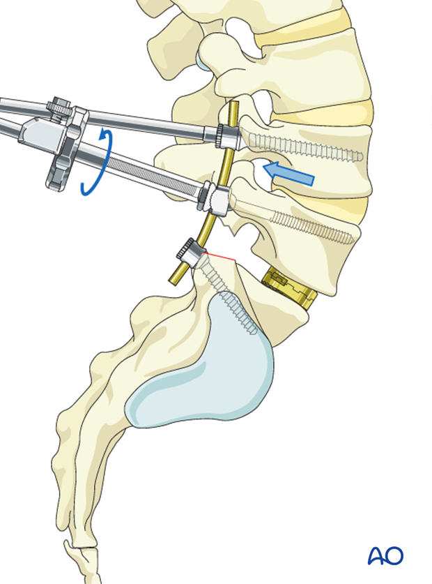
When reduction is satisfactory, all clamps are tightened.
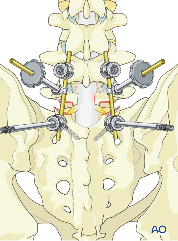
The reduction sleeve is removed,
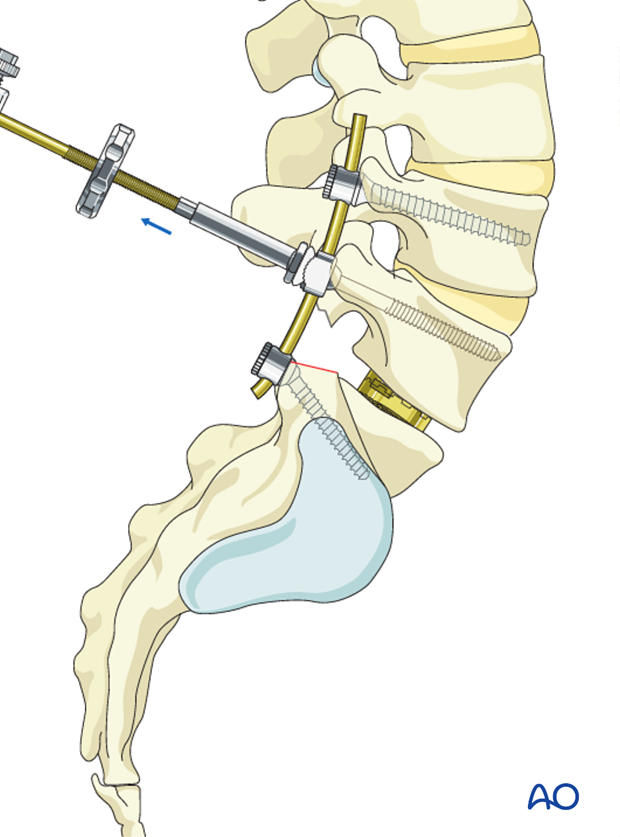
The Schanz pins are cut to the appropriate length.
6. Alternatives
The following alternatives also exist:
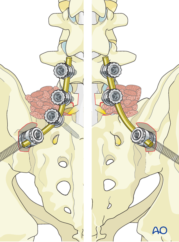
- L4-L5-S1-Ala
- L4-L5-Ala
7. Posterior lateral fusion
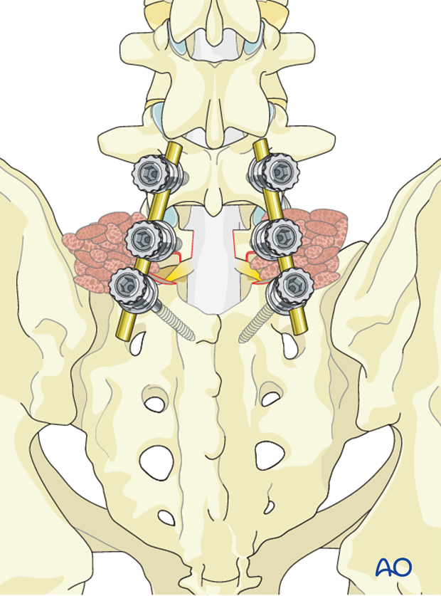
Posterior lateral fusion should be carried out across the transverse process of L4 to sacral ala remove S1 (or the - remove sacral ala. if included in the fixation).
Bone graft is impacted in the lateral gutters.
8. Aftercare
Detailed postoperative neural assessment must be conducted, specifically looking at the integrity of the L5 nerve root as well as sacral nerves controlling bowel and bladder.
Patients with high grade spondylolisthesis that have been reduced are at high risk of postoperative foot drops secondary to neuro traction injury. To minimize such injuries patients are immediately placed in bed with knees and hips flexed. As long as no neuro injury can be identified the leg can be gradually extended after day two. If neuro injury can be identified, the period of flexion should be extended.
Patients are made to sit up in the bed on the first day after surgery. Bracing is optional. Patients with intact neurological status are made to stand and walk on the second day after surgery. Patients can be discharged when medically stable or sent to a rehabilitation center if further care is necessary. This depends on the comfort levels and presence of other associated injuries.
Patients are generally followed with periodical x-rays at 6 weeks, 3 months, 6 months, and 1 year looking for spinal fusion.
Patients with a diagnosis of dysplastic spondylolisthesis run a higher risk of cauda equina and require closer monitoring of their neurological status during and after surgery.


