Anterior open
1. Introduction
The aims of surgery are to:
- improve the spinal curve,
- improve the three dimensional alignment of the spine,
- prevent progression of the curve in the future
- improve cosmesis
- reduce pain
- optimize pulmonary function
- maintain neurological integrity.
This is achieved by correction of the deformity and creation of a solid arthrodesis of the deformed part of the spine.
To illustrate this procedure, we will use a right thoracic curve.
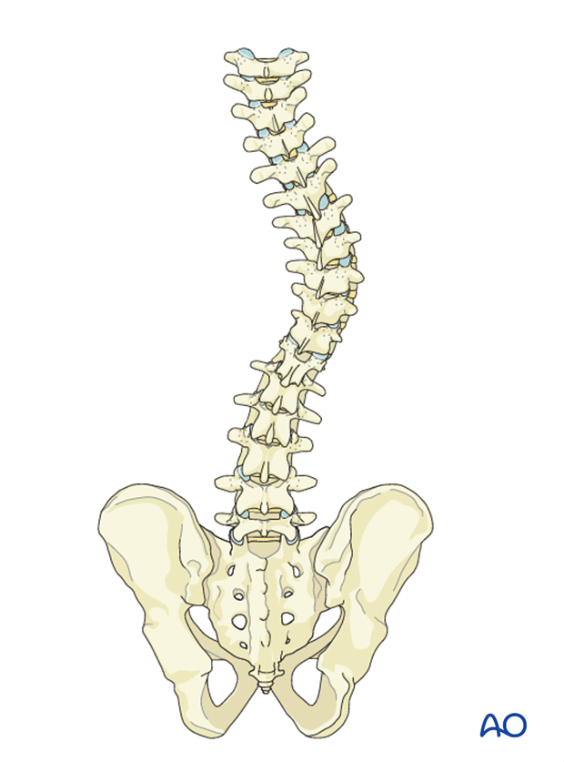
2. Approach and preparation
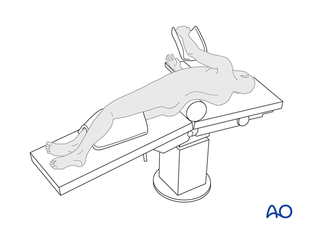
The procedure is performed with the patient in lateral decubitus through one of the folloiwng approaches:
3. Anterior release
The anterior release consists of a thorough discectomy and resection of annulus of the apical intervertebral discs within the proposed instrumented area of the spine. Generally a minimum of 3-4 apical intervertebral discs are excised.
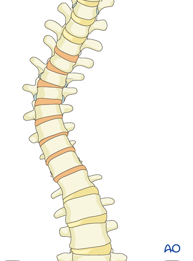
The annulus is incised from the lateral aspect of the spine with a scalpel. The disc is removed using curettes and rongeurs.
Removal of the posterior annulus is optional.
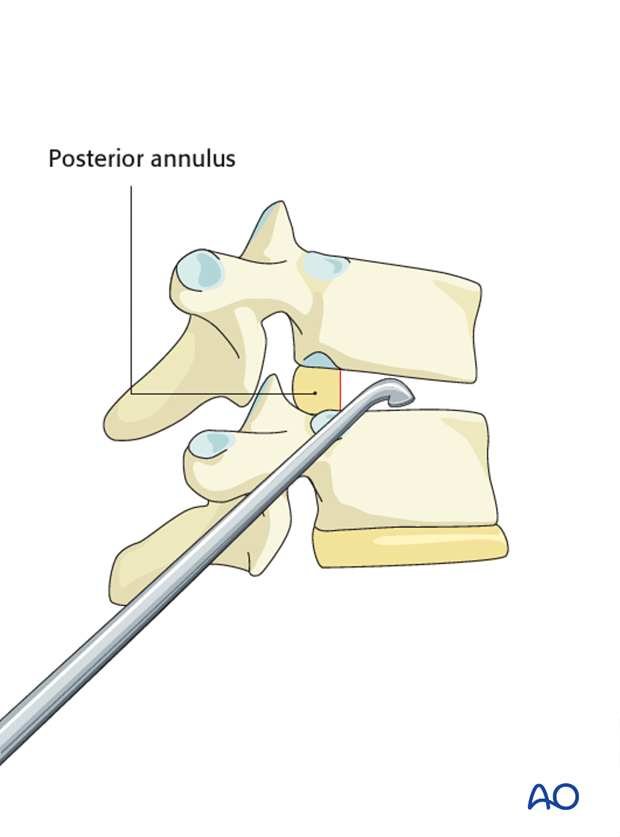
The anterior longitudinal ligament is usually not cut during this procedure and helps to protect the instruments from damaging the anterior vessels.
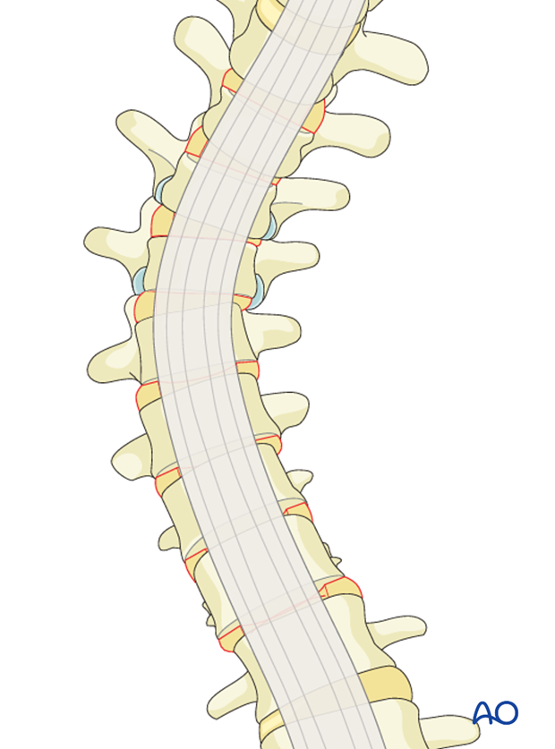
Removal of the posterior annulus
Due to limitation of visualization, care should be taken to excise the posterior annulus if deemed necessary. Excision of the rib head will further improve flexibility and aid in the exposure and excision of the posterior annulus.
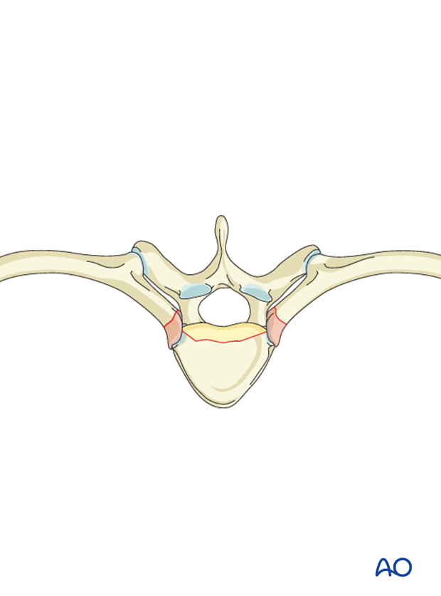
Excision of cartilaginous end plates
The cartilaginous end plate is excised using a curette, a periosteal elevator or a chisel in order to reduce the risk of pseudoarthrosis.
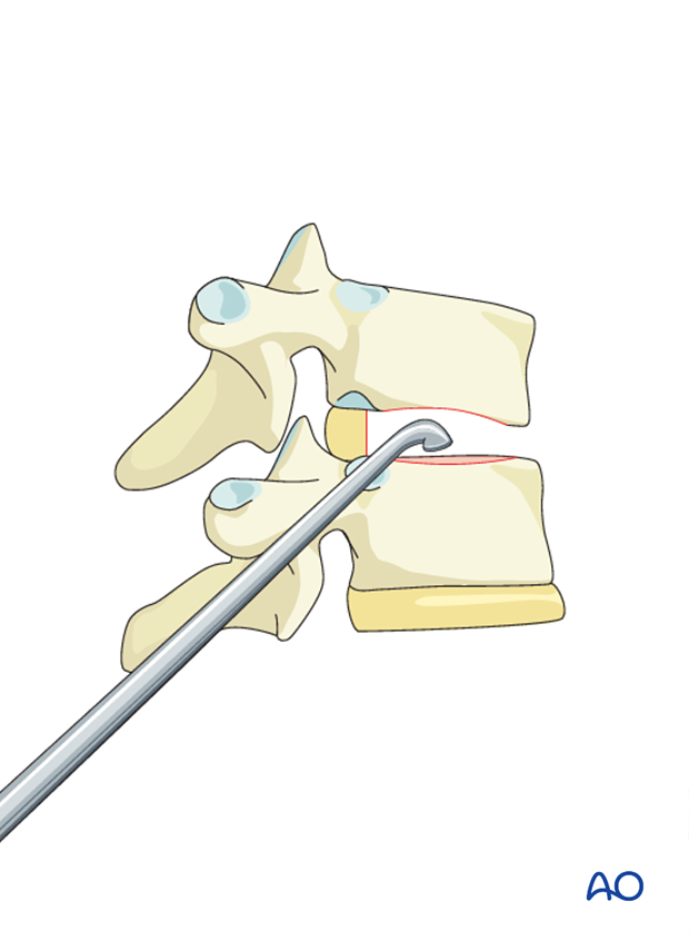
4. Instrumentation
Generally the curve is instrumented from end vertebra to end vertebra, with two bicortical screws at every level. Further details on selection of fusion level can be found here.
A double rod construct is preferred because of increased rotational stability. However, the size of the vertebral bodies will need to be taken into account. For smaller patients, it may not be possible to use two rods.
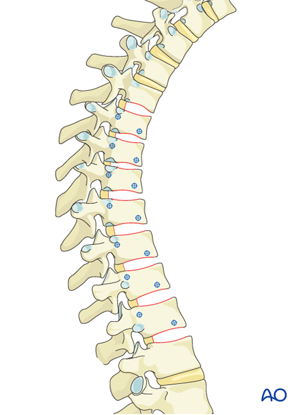
Screw placement
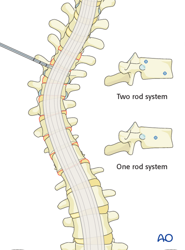
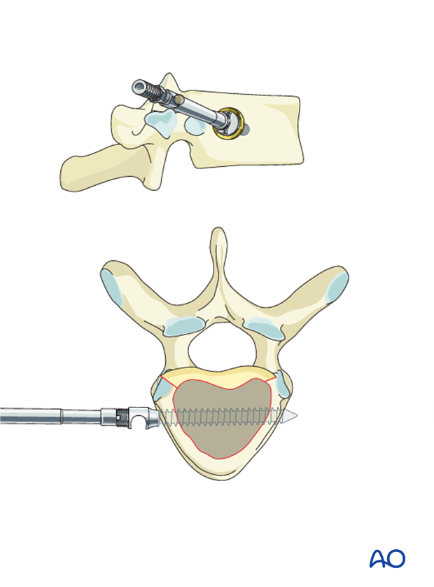
For two-rod constructs one screw is placed as posteriorly as possible after removal of the rib head.
For a single rod construct, the screw should be placed midway between the anterior and posterior margins of the vertebral body, and is aided by removal of the rib head, although this is not always necessary.
The screw holes are prepared using an awl and a probe. Generally a drill is not required. The screws are inserted transversely from one lateral cortex to the contralateral cortex under image intensifier control, or by direct palpation of the contralateral cortex.
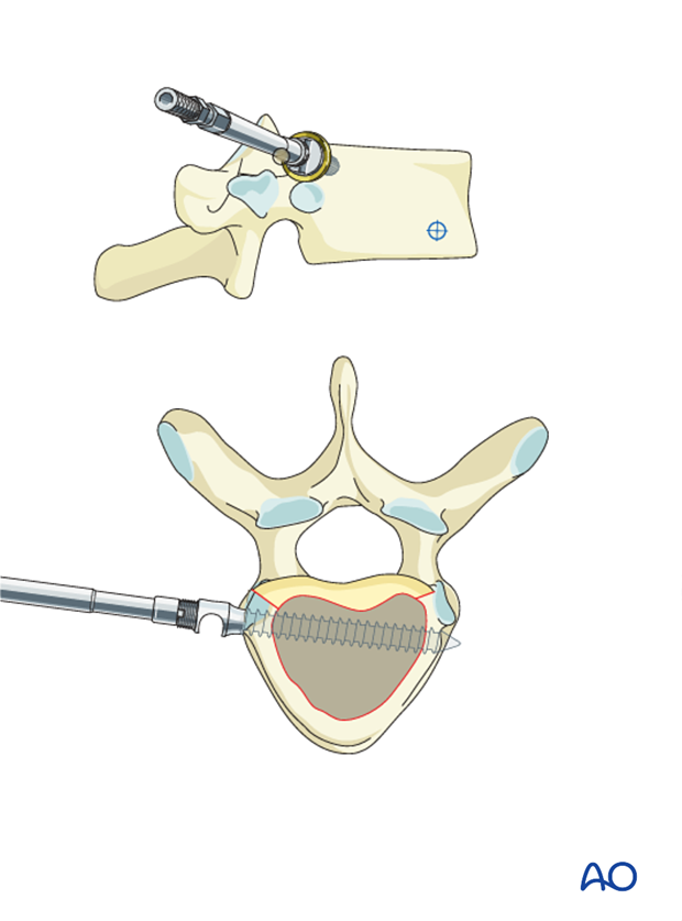
Posterior screw
In the thoracic spine 1 cm of rib head will need to be resected with a rongeur to be able to insert the posterior screw.
The posterior screw is aimed slightly anteriorly to avoid the spinal canal.
If the posterior annulus has previously been removed, the spinal canal can readily be visualized thus ensuring correct placement of the screw.
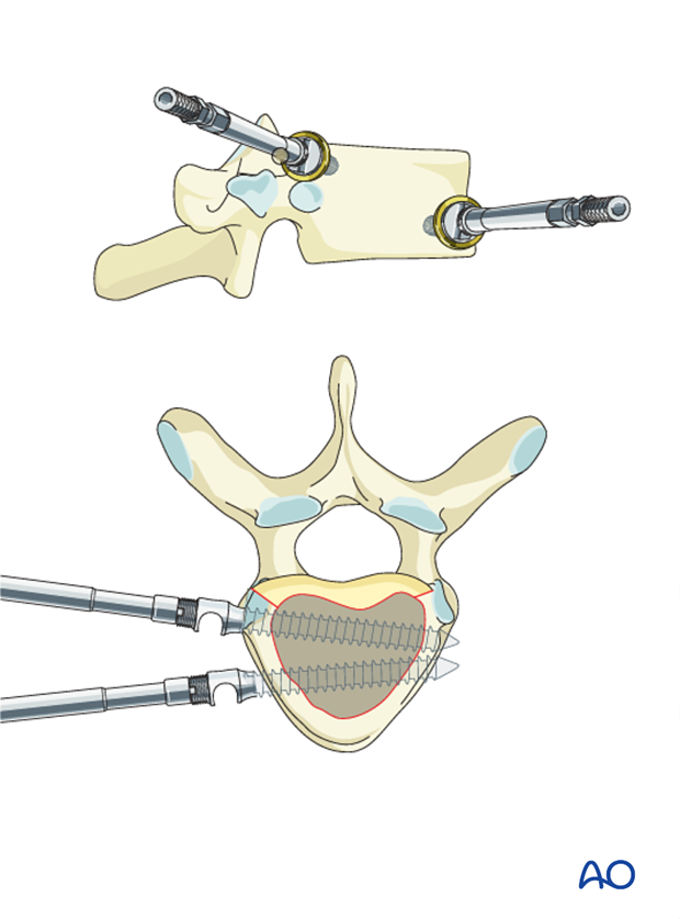
Anterior screw
The anterior screw is aimed slightly posteriorly. In this way the screws are triangulated and the pullout strength is improved.
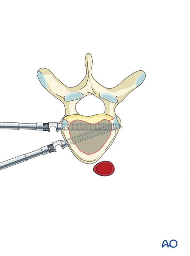
Pitfall: A too anterior placement of the anterior screw may impinge on the vessels and lead to (late) damage to the aorta which is continuously pulsating.
This could be related to a too anterior insertion of the posterior screw eg. if the rib head has not been resected.
Single-screw placement
1 cm of rib head can be resected with a rongeur to facilitate screw insertion.
The screw is directed parallel to the posterior annulus and is aimed to exit the opposite cortex at the mid circumference of the vertebral body.
If the posterior annulus has previously been removed, the spinal canal can readily be visualized thus ensuring correct placement of the screw.
Arthrodesis
The arthrodesis (spine fusion) is achieved through an interbody fusion.
Morselized rib graft supplemented with bone substitutes or iliac crest bone graft is placed in the interbody space.
For Lenke 1curves with a negative sagittal modifier (hypo kyphosis),
interbody cages are not required and bone on bone endplate contact with limited interbody morsalized bone graft is sufficient. By not using structural grafts or cages it is possible to compress the vertebra on to each other, thereby enhancing the thoracic kyphosis.
For Lenke 1 with a normal sagittal modifier (normal kyphosis), some interbody grafting may be required to prevent postoperative hyper kyphosis.

5. Correction of the deformity
The discectomy shortens the anterior column of the spine and makes the motion segments more mobile.
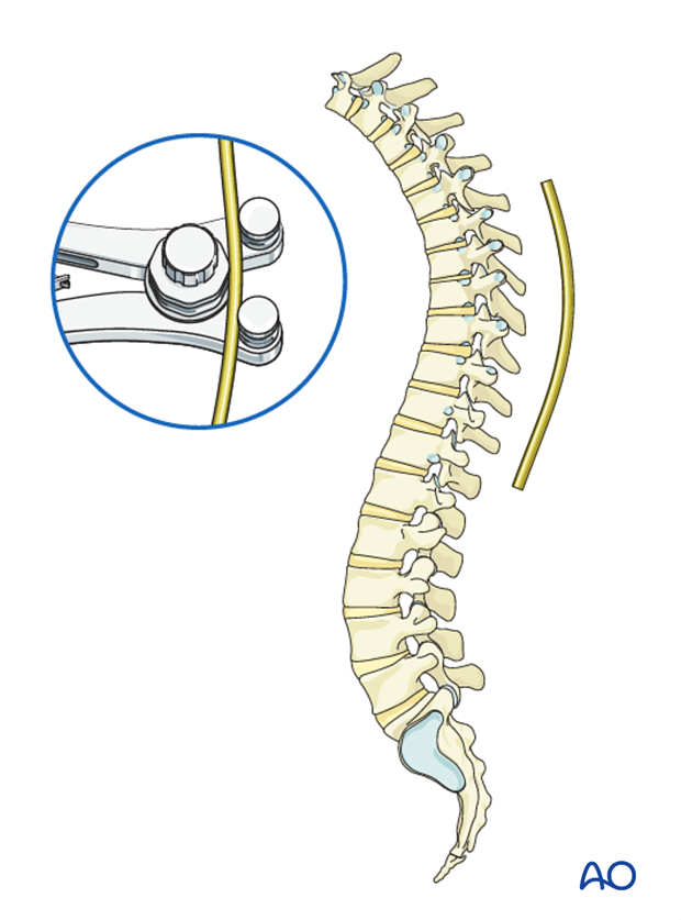
Rod bending
The rods needs to be bent into the correct sagittal profile, which means a mild thoracic kyphosis.
Rod insertion
First correction can be achieved by introducing one or both rods, locking the rods proximally in the screw heads and cantilevering the rods down into each subsequent level below.
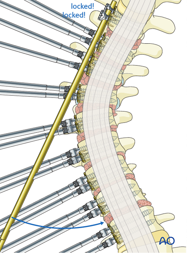
Pitfall: In case a single screw has been used in the UIV, it is best to lock the rod into the proximal two vertebrae before starting to canter lever, otherwise the proximal screw may pull out.
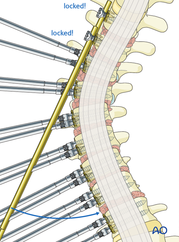
Compression
Second step consists of sequential compression across each disk space, and locking of the rods in each screw head, thereby improving correction and placing the interbody graft under compression.
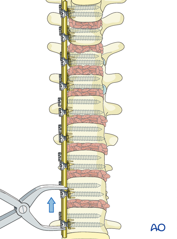
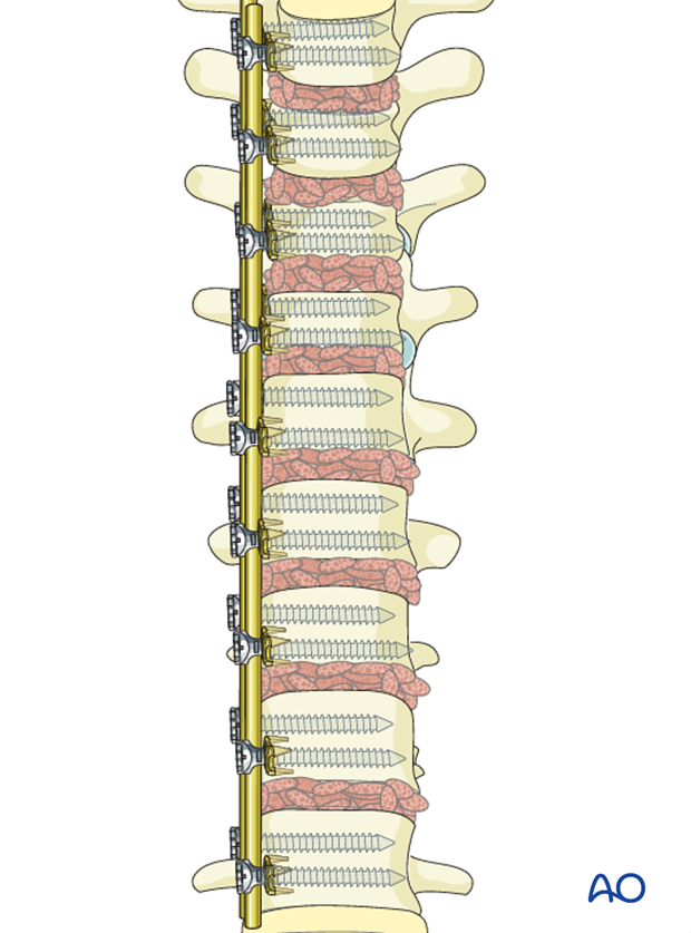
After the correction is completed, the rod will usually be a little longer than necessary.
This can be avoided by anticipating the necessary final length of the rod. At the beginning of the compression, the rod will not reach the last screw, but will do so after all the levels are compressed.
6. Aftercare following correction of spinal deformity
Immediate postoperative medication
Intravenous antibiotics are administrated for at least 24 hours, depending on hospital protocol. The use of an epidural pain catheter vs. intravenous patient controlled analgesia (PCA) are utilized for acute pain management.
Mobilization
Early mobilization out of bed, is preferably started the day after surgery. Generally a postoperative brace/orthosis is not required.
Postoperative imaging
It is appropriate to obtain upright PA and Lateral xrays of the patient at some point early postoperative either before the patient is discharged from the hospital or at the 1st postoperative visit as an outpatient
Restriction of activities
To allow the bone to heal and form a solid arthrodesis, some restriction of sports activities, especially contact sports, is usually advised for 6 months.
Postoperative complications
Early postoperative complications include:
- Postoperative wound infection
- Urinary tract infection
- Respiratory complications such as pneumonia
Late postoperative complications include:
- Pseudarthrosis with loss of correction
- Late deep wound infections
- "Adding on" which is progression of scoliotic deformity in the non-instrumented spine.
- Crankshaft phenomenon (progression of scoliotic deformity within the instrumented spine).
- Implant failure or other implant related complications
In the long term adjacent segment degenerations above and below the instrumented spine may occur.


