ORIF – plate fixation
1. Introduction
The reason to fix this group of fractures is to realign the lateral column. This achieves:
- Optimal rotator cuff function
- Prevention of obstruction of external rotation by posterior impingement
A more detailed description of this procedure is provided in the AO Surgery Reference adult trauma Scapula module.
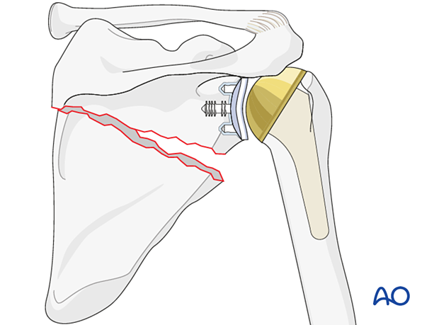
Plate options
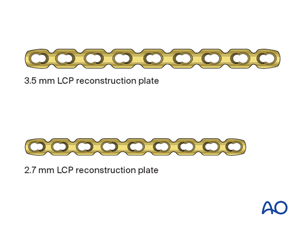
Different types of plate can be used depending on bone quality, fracture location, and surgeon preference:
- 3.5 mm LCP reconstruction plate (suggested for osteoporotic cases)
- 2.7 mm LCP reconstruction plate (suggested for osteoporotic cases)
2. Patient preparation
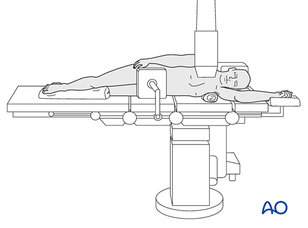
This procedure may be performed with the patient in a lateral decubitus position.
Patient positioning should be discussed with the anesthetist.

3. Approach
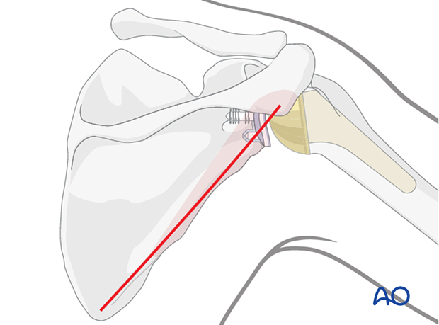
Plating of the glenoid neck and scapula is performed through a posterior approach to the scapular body.
4. Reduction and fixation
Reduction
The fracture can be reduced directly using clamps and indirectly using arm position.
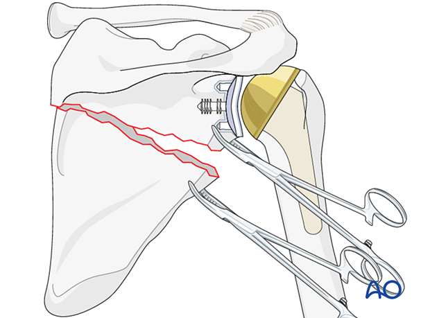
Plate position and contouring
Preoperative planning is crucial to determine optimal plate length and position.
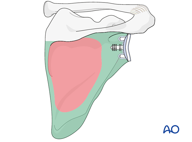
Regions of optimal bone thickness appropriate for plate fixation are shown in green in this illustration.
The lateral border of the scapular is narrow. The angle between the lateral border and the glenoid fossa varies but is nearly always between 30° and 45°. In-plane bending of a lateral column plate will be required.
In common with all periarticular fractures, the fixation of the articular segment is prioritized. Sequential fixation to the lateral column follows to achieve a balanced construct.
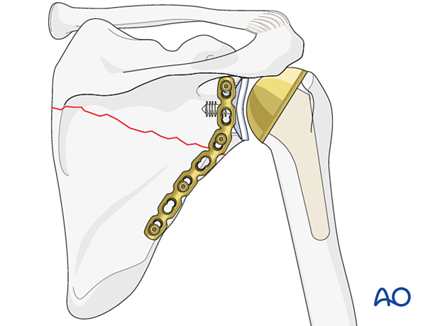
Correct reduction and fixation is verified by image intensification.
5. Aftercare
Postoperative phases
The aftercare can be divided into four phases of healing:
- Inflammatory phase (week 1–3)
- Early repair phase (week 4–6)
- Late repair and early tissue remodeling phase (week 7–12)
- Remodeling and reintegration phase (week 13 onwards)
Full details on each phase can be found here.


