ORIF - Lag screw fixation
1. Introduction
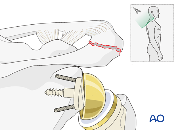
The most common cause of periprosthetic acromial fractures is:
- excess tension on the deltoid in patients with osteopenic bone (typically older female)
These can also be caused by:
- Acute trauma to the shoulder
More information on acromial fractures can be found in the relevant Surgery Reference section.
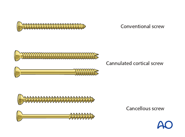
Screw types
Lag screws are used to achieve compression and can be used alone or in combination.
The following screws are useful:
- Conventional 3.5 mm screws
- Cannulated cortical 3.5 mm screws (fully or partially threaded)
- Cancellous 4.0 mm screws (fully or partially threaded)
2. Patient preparation
The standard patient position is the beach chair position with inclination of about 30°. An arm holder may be helpful but is not essential.
Intraoperative fluoroscopy can be helpful.
This procedure can also be performed with the patient in the lateral decubitus position.
Patient positioning should be discussed with the anesthetist.
3. Approach
For fractures of the acromion, a superior scapular approach or a superior acromial anterior to posterior approach (Sabercut approach), is recommended.
4. Reduction and fixation
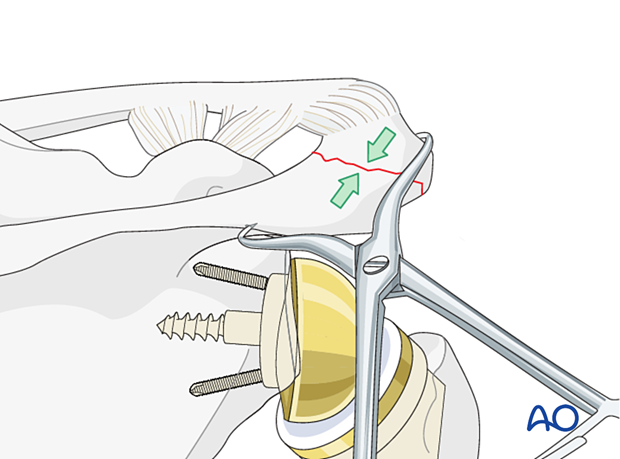
Reduction
Reduction is best achieved using a reduction clamp.
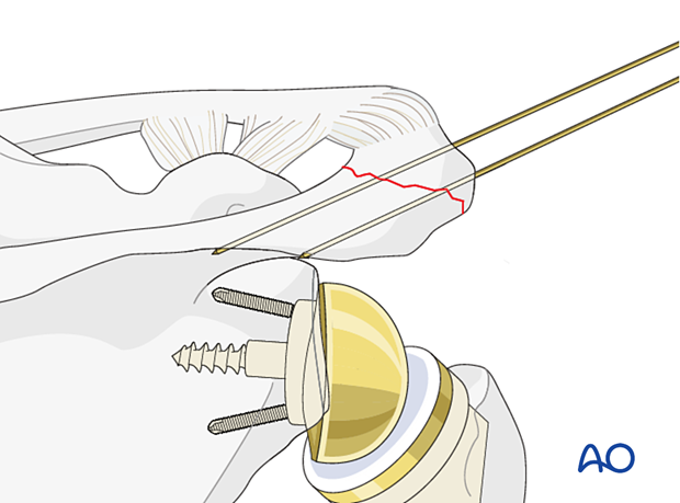
Guide wire insertion/Temporary fixation
At least two screws should be inserted to fix the fracture. These screws should be positioned parallel to each other to optimize compression.
Insert the first guide wire in the desired cannulated screw position. The second guide wire is inserted with the use of the parallel drill guide. The positions of the wires are checked with the image intensifier to make certain that they are in bone and have not exited subacromially and entered the rotator cuff, the supraspinous fossa, or the shoulder joint.
Cannulated screw insertion
Predrill the proximal cortex of the acromion to create two gliding holes. Use 3.5 mm fully threaded cannulated screws of the correct length as lag screws. Washers may be used under the screw heads in osteopenic bone.
Remove the guide wires.
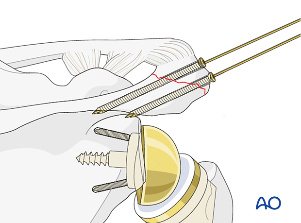
In the setting of an acromial fracture associated with a reverse total shoulder replacement the forces acting on the acromion fragment are greater. In this setting augmented multiplanar fixation is recommended.
Options include:
- Lag screw fixation with cerclage compression wiring
- Dual plate fixation
The cerclage compression wiring fixation for this injury is shown here.
Pearl: It may be impossible to reduce the fracture due to the tension of the deltoid muscle. In this situation options include:
- Single stage - exchange by downsizing of the glenosphere and humeral liner if possible
- Staged surgery - temporary removal of the articulating components to permit healing and then reimplantation
Check the reduction and temporary fixation with an image intensifier.

5. Aftercare
Postoperative phases
The aftercare can be divided into four phases of healing:
- Inflammatory phase (week 1–3)
- Early repair phase (week 4–6)
- Late repair and early tissue remodeling phase (week 7–12)
- Remodeling and reintegration phase (week 13 onwards)
Full details on each phase can be found here.


