ORIF - Cerclage compression wiring
1. Introduction
Most isolated anterolateral acromial fractures can be managed non-operatively.
With a reverse prosthesis, there may be more tension on the deltoid muscle due to lengthening of the arm. Acromial fractures may therefore occur either perioperatively, or after some delay.
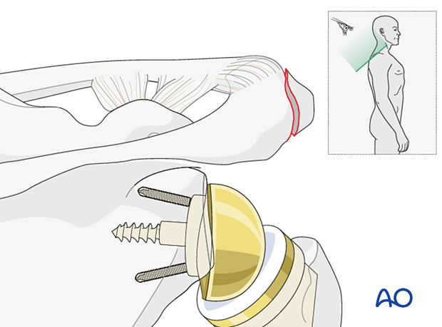
2. Patient preparation
The standard patient position is the beach chair position with inclination of about 30°. An arm holder may be helpful but is not essential.
Intraoperative fluoroscopy can be helpful.
This procedure can also be performed with the patient in the lateral decubitus position.
Patient positioning should be discussed with the anesthetist.
3. Approach
For fractures of the acromion, a superior scapular approach or a superior acromial anterior to posterior approach (Sabercut approach), is recommended.
4. Reduction and fixation
Reduction
Reduction is best achieved using a reduction clamp.
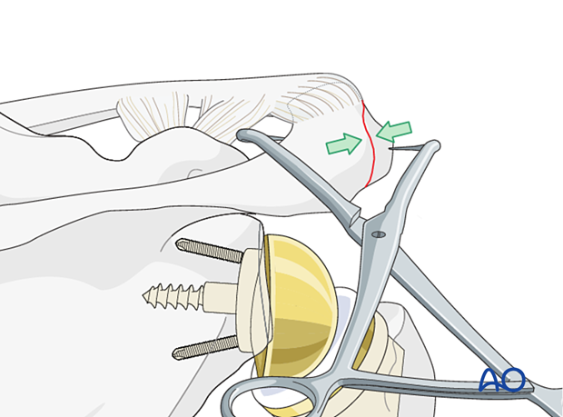
In the setting of an anterolateral acromial fracture associated with a reverse total shoulder replacement the forces acting on the acromion fragment are greater. In this setting augmented multiplanar fixation is recommended. Options include:
- Lag screw fixation with cerclage compression wiring
- Dual plate fixation
The cerclage compression wiring fixation for this injury is illustrated here.
This cerclage compression construct was previously called a “tension band”. More information about the tension band principle can be found here.
K-wire insertion
In an anterolateral fracture with a small acromial fragment, a tension band alone may be sufficient.
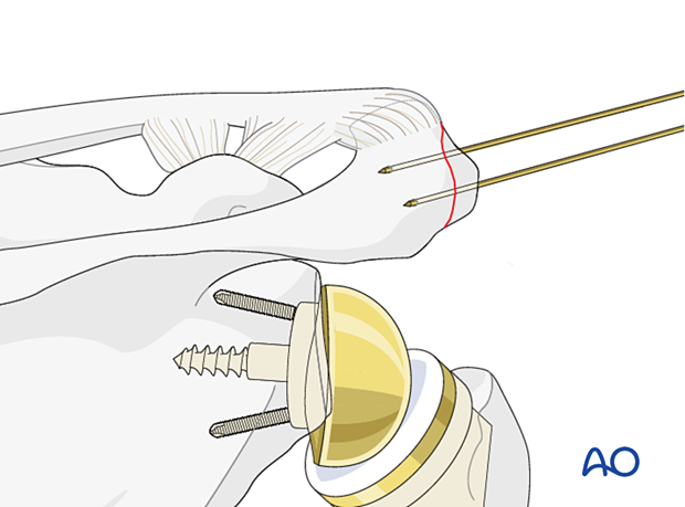
A larger acromial fragment is fixed using a 1.6 mm K-wire inserted from the anterolateral direction.
It should exit the dorsal cortex of the posterior acromion. A second K-wire is inserted parallel to the first with the help of a parallel drill guide. Both K-wires should protrude 5 mm dorsally.
Check the position of the K-wires with an image intensifier to make sure they have not penetrated the subacromial space.
Loop the cerclage wire around the two K-wires as shown. In common with other situations in which cerclage wire is used, form a loop on one limb so that the loop and the wires can be tightened simultaneously. This will allow symmetrical tension to be obtained in both wires.
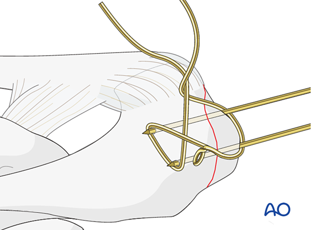
The K-wires are shortened and bent as shown.
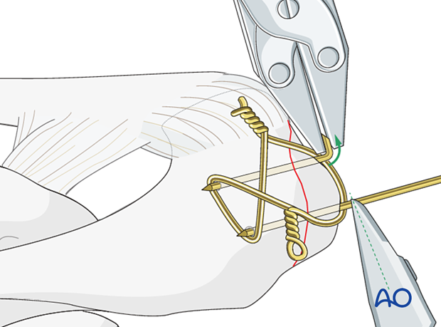
Check the reduction and temporary fixation with an image intensifier.

5. Aftercare
Postoperative phases
The aftercare can be divided into four phases of healing:
- Inflammatory phase (week 1–3)
- Early repair phase (week 4–6)
- Late repair and early tissue remodeling phase (week 7–12)
- Remodeling and reintegration phase (week 13 onwards)
Full details on each phase can be found here.


