Authors of section

Authors

Samy Bouaicha, Stefaan Nijs, Markus Scheibel, David Weatherby

Executive Editor

Simon Lambert

Open all credits

Humeral osteotomy

1. Introduction

If there is bone ongrowth or a well-fixed cement mantle which prevents easy removal of a stem, then a humeral osteotomy may be required.

2. Approach

Skin incision

A deltopectoral approach is performed.
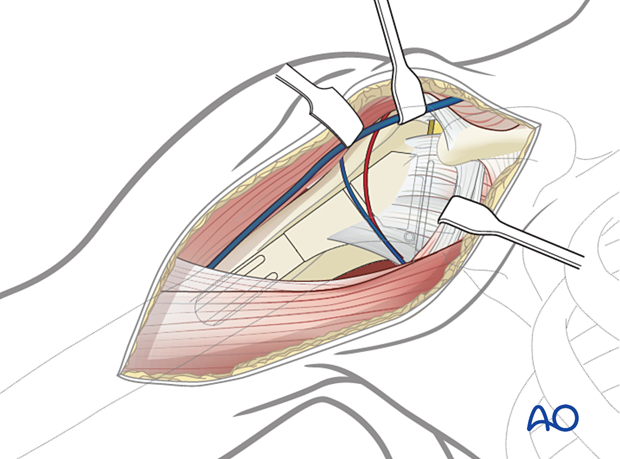
Humeral osteotomy - approach

Deep approach

The following landmarks are used to make the vertical osteotomy:

  • pectoralis tendon
  • deltoid tendon
  • long head of the biceps tendon and bicipital groove
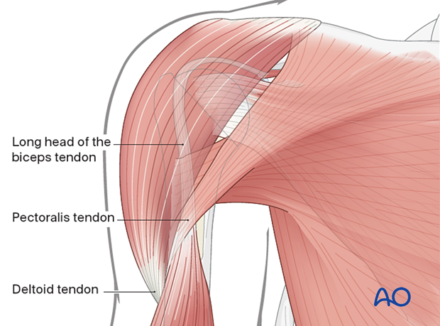
Humeral osteotomy - deep approach

3. Anatomic configuration

A vertical osteotomy is made between the pectoralis and deltoid tendons, from the most proximal extent of the humerus to the distal end of the prosthesis.

Fluoroscopy may be helpful in identifying the level of the distal end of the prosthesis.

Humeral osteotomy - a vertical osteotomy is made between the pectoralis and deltoid tendons

A microsaw is used to cut straight down to the implant.
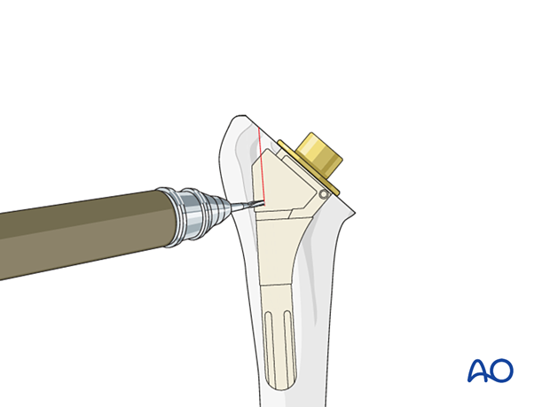
Pearl: If a microsaw is not available the osteotomy can be created by perforation of the shaft using a 2 mm drill and connecting the perforations with osteotomes.
Humeral osteotomy – a microsaw is used to cut straight down to the implant

A single cut is performed from the proximal to the distal aspect of the stem.
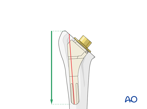
Pearl: A 3.5 mm drill hole can be made at the distal end of the cut to prevent inadvertent propagation of the split into the distal humeral shaft.
Humeral osteotomy – a single cut is performed from the proximal to the distal aspect of the stem

Multiple osteotomes are inserted in the cut to spread it open.
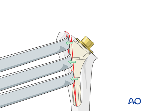
Humeral osteotomy – multiple osteotomes are inserted in the cut to spread it open

The revision extractor instrument is attached to the stem and backslapped in the axis of the humerus.
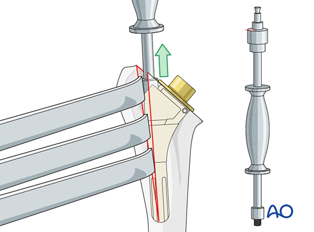
Humeral osteotomy – the revision extractor instrument is attached to the stem and backslapped in the axis of the humerus

If the implant is cemented, and cement remains in the medulla after removal of the implant, the cement is removed with osteotomes, curettes, and rongeurs.
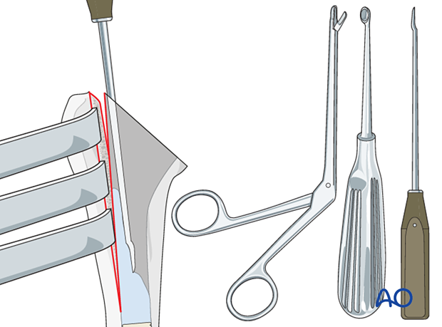
Humeral osteotomy – if the implant is cemented, and cement remains in the medulla after removal of the implant, the cement is removed with osteotomes, curettes, and rongeurs

4. Repair of the osteotomy

The osteotomy is repaired using cerclage wires or cables. At least two cerclage wires or cables should be used to stabilize the humerus.
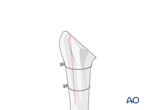
Pitfall: Care must be taken to avoid damaging the axillary and radial nerves during passage of the cerclage wire around the posterior aspect of the humerus.
Humeral osteotomy – the osteotomy is repaired using cerclage wires or cables
Go to diagnosis