Intramedullary nail and plate fixation
1. Principles
Fractures distant from the implant are commonly comminuted in the setting of osteoporotic bone. These fractures can be treated similarly to a femur without prosthesis, with attention towards proximal fixation.
It is important to review prior radiographs of the total hip replacement to evaluate whether or not the stem is stable.
2. Approach
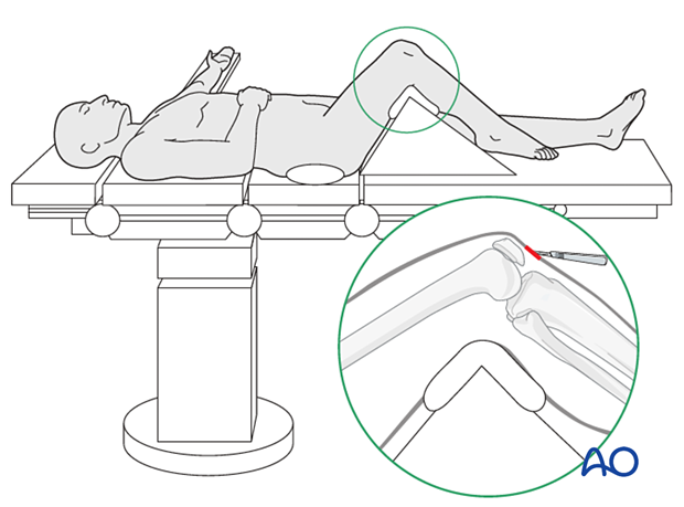
The limb is prepared on a lower extremity radiolucent triangle, and an incision is made over the patellar tendon after indirect reduction.
Further details about the approach for retrograde nailing may be found here.
3. Indirect reduction
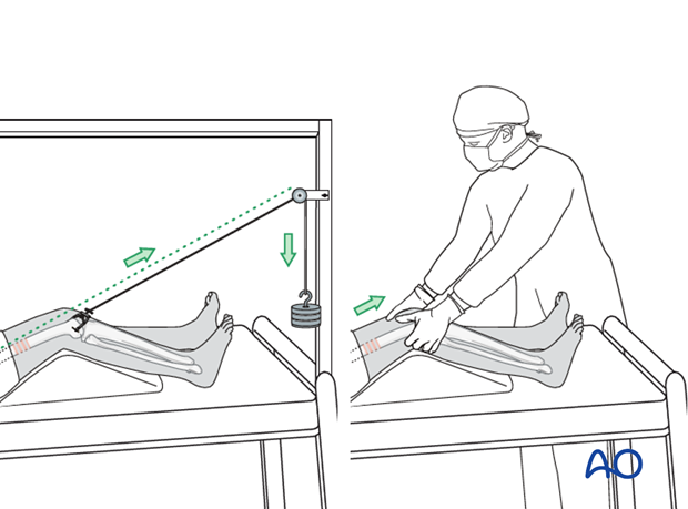
Distal femoral skeleton traction or table traction is critically important for restoration of limb length and rotation.
Reduction techniques
Adjunctive methods of reduction include the utilization of a large femoral distractor, or Schanz pin manipulation of fracture fragments.

Reduction tools include percutaneous bone hooks, ball spike pusher, and Hohmann retractors.
For additional details about indirect reduction please refer to the femur shaft section of the AO Surgery Reference.
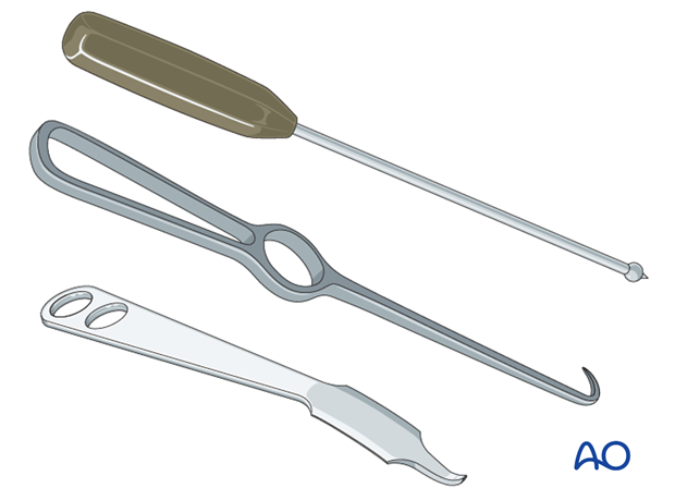
Reduction of comminuted supracondylar femur fractures can be exceptionally difficult to obtain and maintain.
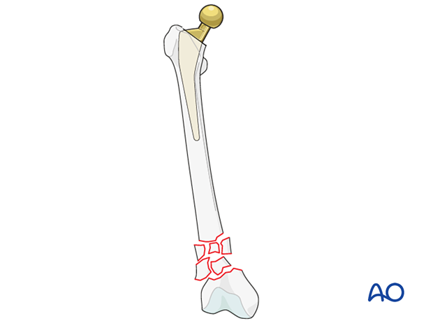
This is complicated by the need to span the entirety of the femur because of the total hip replacement.
Careful attention must be given to both coronal and sagittal plane alignment.
4. Implant considerations
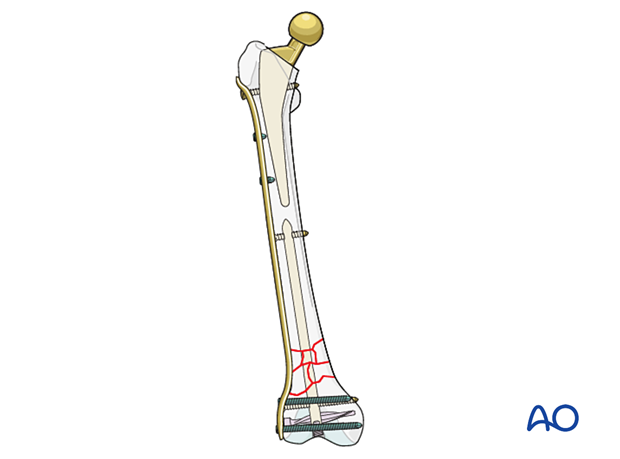
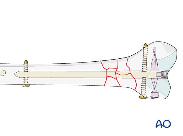
Given the complexity of healing of Vancouver C comminuted supracondylar fractures, it is possible to utilize both an intramedullary nail in a retrograde fashion, along with lateral plating support of the fracture. This can allow for earlier mobilization.
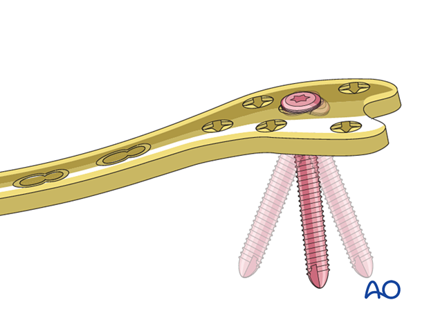
To achieve fixation around the intramedullary nail at the knee, a variable angle locking plate might be beneficial.
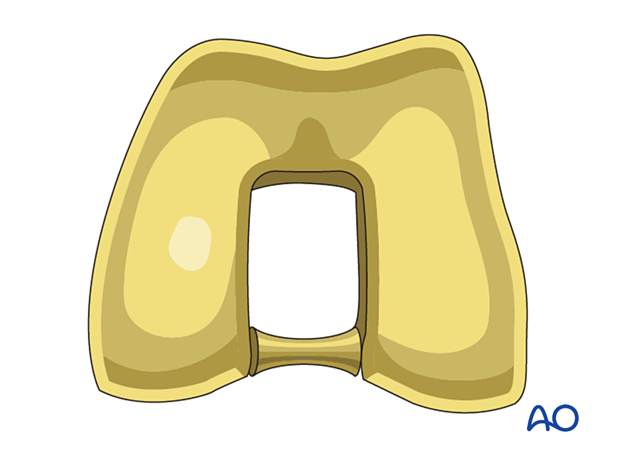
It is recommended that a replacement polyethylene component for the knee is available if damage should occur to the existing polyethylene.
5. Intramedullary nail application
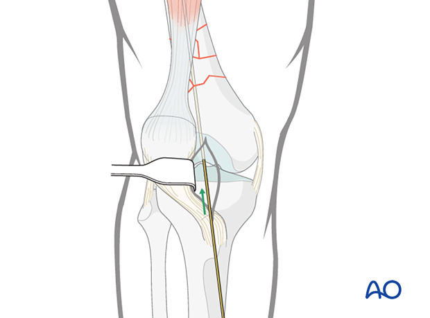
Opening the medullary canal
A guide wire is introduced into the distal femur segment in standard retrograde nail fashion.
The distal femur is opened utilizing a cannulated drill bit of appropriate size.
Reaming
The guide wire and the drill bit are removed, and a ball tipped reaming wire is introduced across the proximal segment, the fracture site and up to the distal tip of the femoral prosthesis.
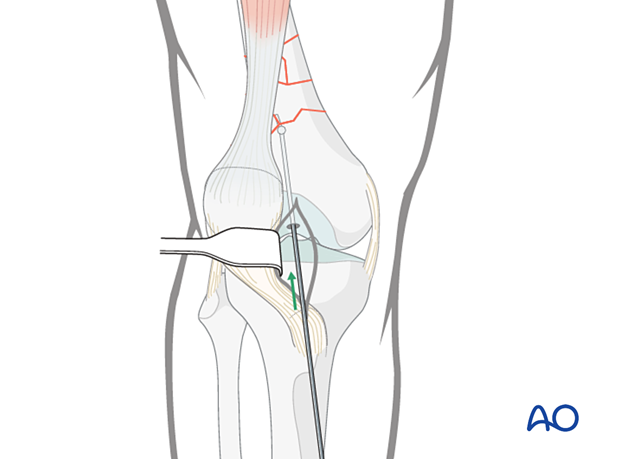
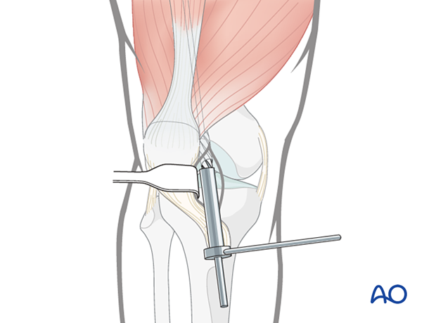
The femur is prepared performing sequential reaming.
Nail insertion
The intramedullary nail is inserted over the guide wire into the distal fragment, to abut the femoral prosthesis.
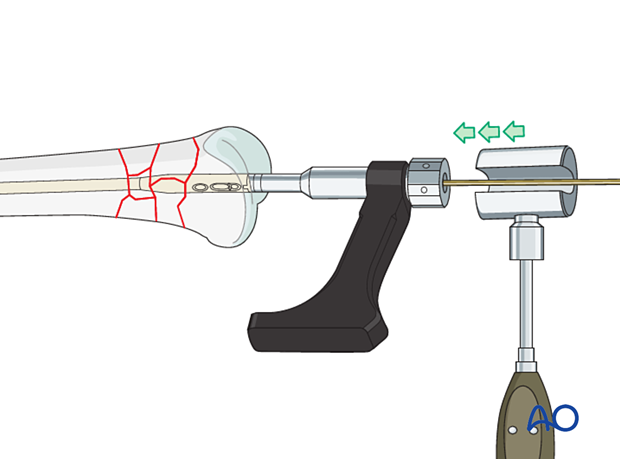
The nail is then interlocked proximally and distally utilizing standard techniques.
For more details regarding retrograde femoral nailing, depending on the fracture pattern, please refer to the dedicated section of the distal femur in the AO Surgery Reference.
6. Lateral plate application
A supplemental plate is applied to the lateral aspect of the femur.
For more details regarding condylar plating please refer to the dedicated section of the distal femur in the AO Surgery Reference.
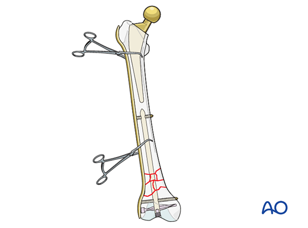
The plate is secured to the femur using hybrid fixation methods.

Options for additional stability
Additional stabilization around the femoral stem may be obtained with adjunct plate options, such as:
- Unicortical screw fixation
- Cerclage cables integrated into the plate
- Locking attachment plate

7. Aftercare following ORIF
Postoperative management
Postoperative management should include careful monitoring of hematocrit and electrolytes particularly in the elderly patients.
Postoperative IV antibiotics should be administered up to 24 hours.
Consideration should be given to anticoagulation for a minimal course of 35 days. If there are thromboembolic complication this treatment is extended.
Drains can be discontinued when output is less than 30 to 50 cc per 12 hours.
Patient mobilization
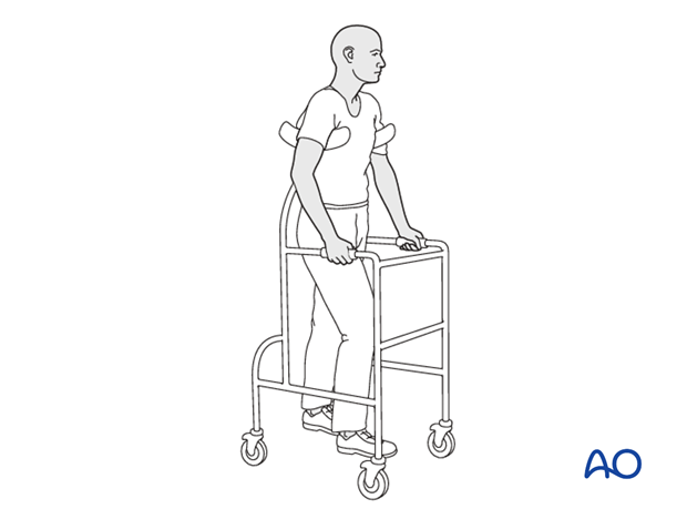
Immediate mobilization of the patient should commence. If fracture stability will allow, the patient should be made weight bearing as tolerated as soon as possible. Long periods of limited weight bearing are extremely detrimental to patient recovery.
Combined constructs of intramedullary nail and plate theoretically provide a load sharing environment, which is conducive to early weight bearing.
Wound healing
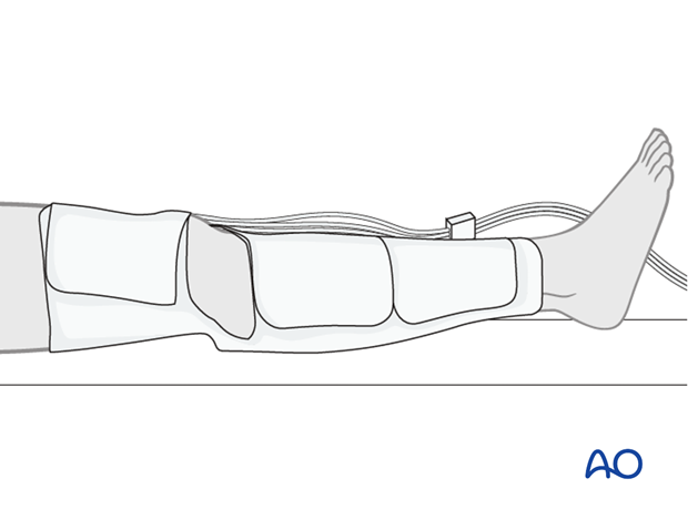
Avoidance of edema postoperatively is critical for both wound healing and patient mobilization. This can be aided by pneumatic compression devices. If negative pressure wound therapy is utilized, it can be discontinued after 5 to 7 days. Staples or sutures are typically removed at 14 to 21 days.
