Complications and technical failures
1. Introduction
Proximal tibial fractures are associated with significant but predictable complications.
Treatment related complications can be minimized by anticipating them and paying attention to the general principles of fracture management.
2. General considerations
The complications associated with proximal tibial fractures include:
- Neurovascular injury
- Compartment syndrome
- Loss of reduction
- Infection
- Cast complications
- Malunion
- Growth disturbance
- Posttraumatic arthritis
3. Neurovascular injuries
Neurovascular injuries may result from the initial trauma, displacement of fracture fragments and secondary (iatrogenic) trauma during management.
Examples include:
- High-energy injuries
- Posterior displacement of an epiphyseal fragment into the popliteal fossa
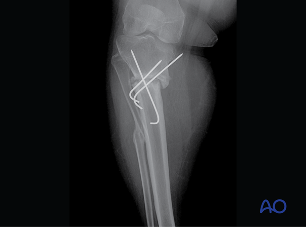
- Penetration with wires/screws
- Pressure from cast/splint over the common peroneal nerve
Early diagnosis and management are crucial to minimize permanent injury.
4. Compartment syndrome
Compartment syndrome is most common in the lower leg.
The diagnosis of compartment syndrome should be suspected in a conscious and alert patient, when the following early signs are present:
- Pain, unrelated to the posture of the limb, made worse by passive movement of muscles in the relevant compartment
- Paresthesia
- Progressive loss of light touch sensation
5. Loss of reduction
Loss of reduction may result from:
- Unstable injury
- Inadequate fixation
6. Infection
The risk of infection is increased in:
- Open fractures and associated soft-tissue injuries
- Soft-tissue tethering around K-wires, pins, or prominent implants
- Delayed removal of exposed K-wires
7. Cast complications
Pediatric fractures are commonly treated with a cast, but this requires close attention to detail.
Preventive measures during application:
- Padding all pressure points (eg, malleoli, patella, fibular head)
- Correct application of cast bandage
- Avoid application of a complete cast in a swollen leg
- Exposure of all toes to allow assessment of distal circulation
Preventive measures after cast application:
- Elevation of the leg
- Instructions to care givers:
- Check toe perfusion
- Check for excessive swelling
- Avoid insertion of objects between cast and skin
- Avoid wetting the cast and padding
Preventive measures during cast removal:
- Using sharp saw blades to avoid injury
8. Delayed union and nonunion
Nonunion is rare in pediatric tibial shaft fractures and requires investigation for a pathological cause.
Delayed union may occur as a consequence of:
- Instability
- Persistent fracture gap
- Open reduction
- Inappropriate choice of implant
- Severe soft-tissue damage
- Infection
- Devascularization
- Delayed weight bearing
- Bone loss
- Vitamin D deficiency

9. Malunion
Causes:
- Inadequate reduction
- Inappropriate choice or use of implant
10. Implant pain/prominence
Causes:
- Large plates with inadequate soft-tissue cover
- Long screws
- Prominent nail ends
11. Joint stiffness
Causes:
- Prolonged immobilization due to:
- Pain
- Instability - Extensive soft-tissue injury
12. Peri-implant fracture
Causes:
- Stress riser from plate
- Pathological bone (eg, disuse osteopenia, osteogenesis imperfecta)
13. Growth disturbance
The proximal tibial physis contributes significantly to the growth of the lower limb.
Disturbance of physeal growth may result from the initial injury or from repeated manipulation.
This may result in premature arrest of the entire physis which causes shortening or partial arrest, which causes progressive deformity.
Prevention by avoiding further injury to the physis during fracture management by:
- Anatomical reduction
- Gentle manipulation
- Limiting attempts at reduction
- Avoiding or limiting K-wire insertion across the physis
- Using smooth, narrow K-wires across the physis
Clinical and radiographic surveillance is important to detect growth disturbance, particularly in younger patients. If this is suspected, MRI/CT scan is recommended.
Cozenʼs phenomenon
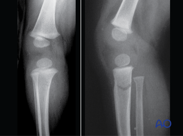
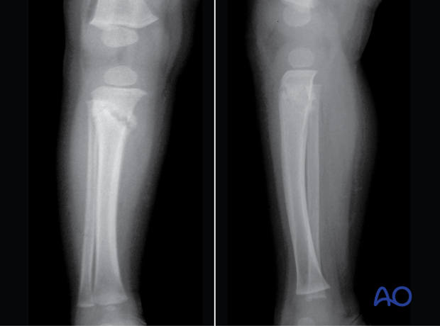
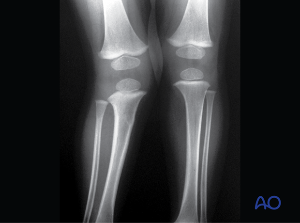
Incomplete proximal tibial metaphyseal fractures in young patients can be associated with progressive apex medial (valgus) angulation (Cozen’s phenomenon). When present, this deformity develops in the months following injury and the majority will remodel within a few years.
The parents/carers should be alerted to this possibility at the first presentation.
These initial x-rays show an incomplete metaphyseal fracture of the proximal tibia in a 3-year-old child.

In these x-rays 3 weeks postinjury, callus formation can be seen.

In this x-ray 18 months postinjury, a valgus (Cozen’s) deformity is visible.

14. Posttraumatic arthritis
Intraarticular (Salter-Harris type-III and -IV) fractures of the proximal tibia are associated with posttraumatic arthritis if anatomical reduction has not been achieved during surgery.


