Closed reduction - K-wire fixation
1. General considerations
This technique is used after open or closed reduction of extraarticular proximal tibial fractures including Salter-Harris I and II, and proximal metaphyseal fractures in patients with an open tibial physis.
These fracture patterns require temporary fixation across the physis to produce adequate stability.
To minimize secondary damage to the physis, manipulation must be gentle.
Multiple passes across the physis with a K-wire should be avoided.
Smooth, narrow K-wires reduce the risk of physeal damage.
Threaded K-wires may increase fracture stability.
Closed vs open reduction
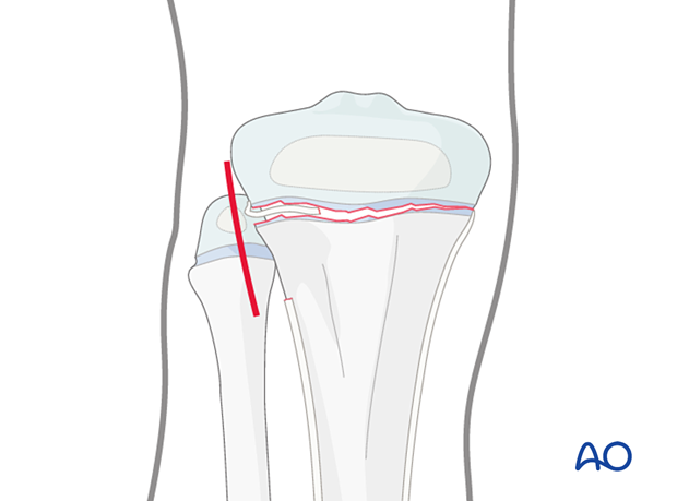
If initial closed reduction is unsuccessful, this is usually due to periosteum entrapped on the side that has failed in tension.
2. Instruments and implants
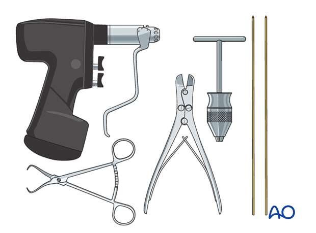
The following equipment is needed:
- K-wires of appropriate sizes
- Drill or a T-handle for manual insertion
- Wire cutting instruments
- Standard orthopedic instrument set
3. Patient preparation
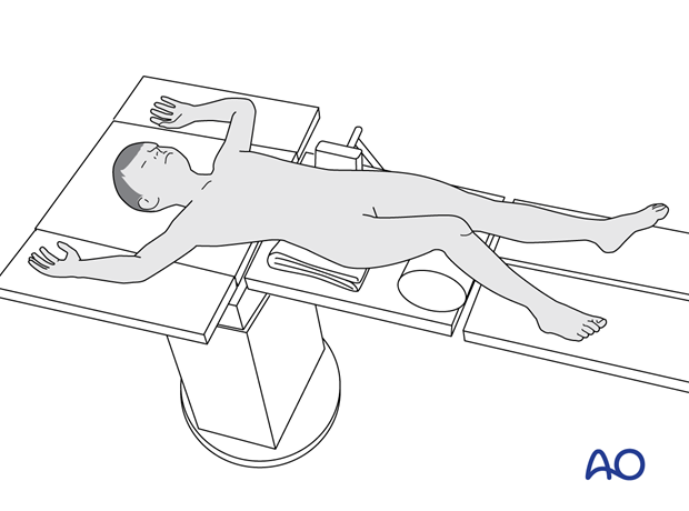
Place the patient in a supine position on a radiolucent table, with a bolster under the ipsilateral flank.
Breaking the table to allow knee flexion may facilitate traction and reduction.
4. Reduction
Gentle, prolonged longitudinal traction is the most important maneuver to avoid damage to the physis.
This should be followed by manipulation of the limb for fracture reduction.
An assistant should be available to apply countertraction.
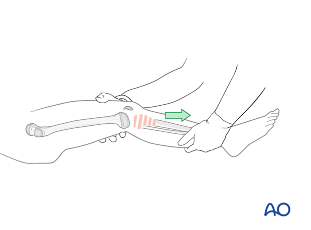
Confirm reduction with an image intensifier.
If a gap remains after reduction, a small open approach may be required to release the periosteum.
Open reduction should be considered if closed reduction is unsuccessful.

5. Fixation
Insertion of K-wires
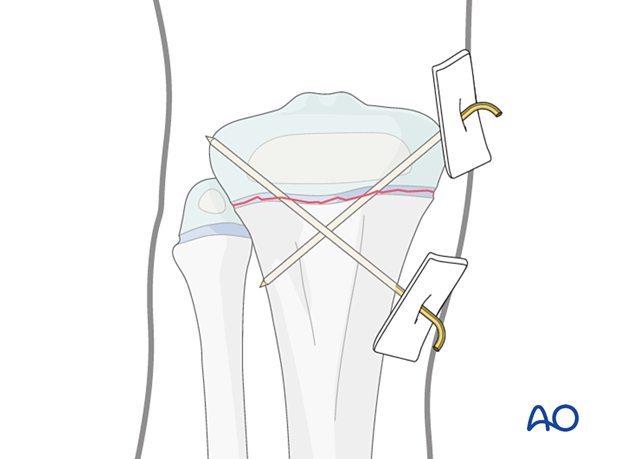
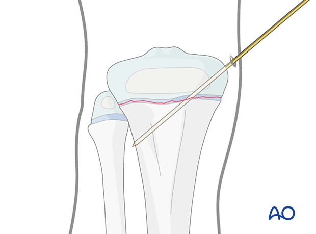
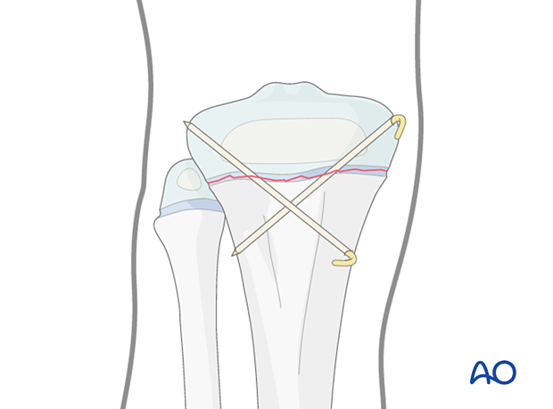
Insert the first K-wire from proximal to distal starting medially.
Advance the K-wire across the physis after confirming a trajectory that will engage the metaphysis of the proximal tibia on AP and lateral views.
Engage the K-wire in the cortical bone of the tibia.
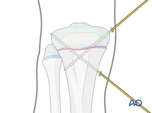
The second K-wire may be inserted from distal to proximal from the medial side to avoid injury to the common peroneal nerve.
Take care not to penetrate the articular surface with the tip of the K-wire.
Alternatively, the K-wire may be inserted from proximal to distal starting laterally taking care not to injure the peroneal nerve.
In either case, maximize the spread of the K-wires at the fracture site to increase stability.
Confirm stability of fracture fixation with an image intensifier.
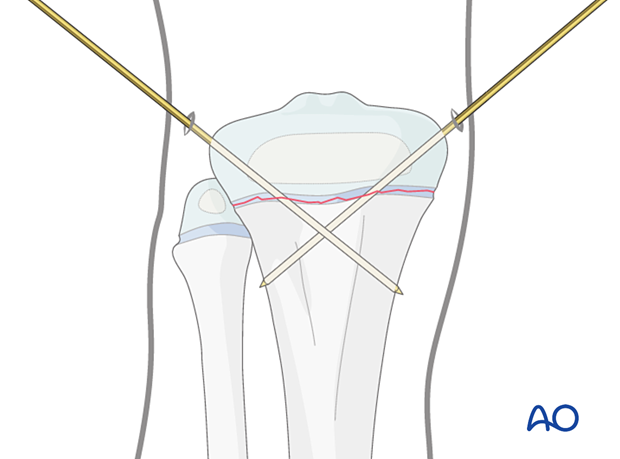
K-wire cutting and dressing
Bend the K-wires approximately 2 cm from the skin to accommodate for swelling as illustrated.
Cut the K-wires and apply a dressing or sponge to stabilize the K-wires against the skin.

K-wires may be buried under the skin, but this requires further surgery for wire removal.
6. Final assessment
Confirm fracture reduction and implant position with an image intensifier.
7. Immobilization
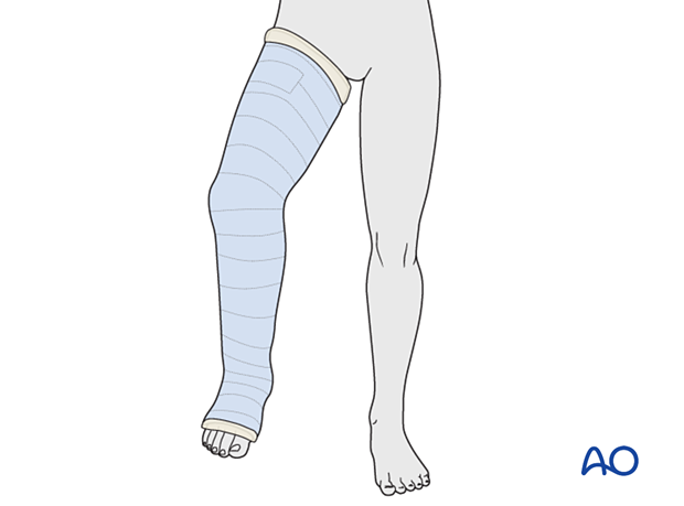
A cast is mandatory because K-wires in isolation are inadequate to neutralize muscle forces.
Apply a long leg cast with the knee flexed to 30°–45° for 3–6 weeks, depending on the age of the patient.
8. Aftercare
Immediate postoperative care
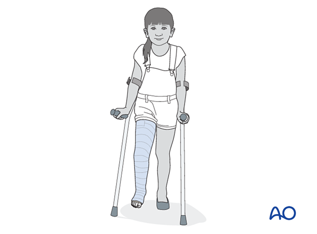
The patient is kept touch-weight bearing.
Older children may be able to use crutches or a walker.
Younger children may require a period of mobilization in a wheelchair.
Neurovascular examination
The patient should be examined frequently in the initial period following the injury, to exclude neurovascular compromise or evolving compartment syndrome.
High-energy fractures are associated with vascular injuries and require careful clinical assessment.
Follow-up, cast and K-wire removal
These fractures heal quickly, and cast and K-wires are typically removed, following an x-ray, 3–6 weeks after injury.
Mobilization
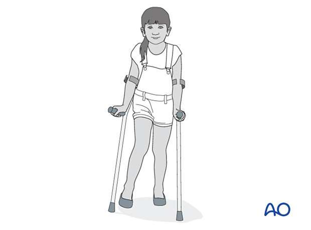
Once K-wires and cast are removed, partial weight-bearing and knee range-of-motion exercises are started with gradual return to normal activity.
Follow-up for growth disturbance
All patients with fractures of the proximal tibia should have clinical and radiological examination 8–12 weeks postoperatively to confirm healing and alignment.
Clinical examination should be repeated at intervals for at least one year to detect early signs of growth disturbance.
If there is a clinically relevant leg length discrepancy or malalignment, radiological assessment is required.


