Proximal humeral anatomy
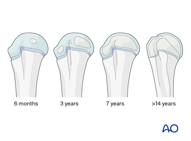
1. Developmental anatomy
An understanding of the development of the proximal humerus is necessary for accurate fracture diagnosis.
The appearance of secondary ossification centers is as follows:
- Proximal humeral epiphysis; 6 months
- Greater tuberosity; 3 years
- Lesser tuberosity; 5 years
Other important radiological events include:
- Coalition of tuberosities; 5–7 years
- Fusion of tuberosities with humeral epiphysis; 7–13 years
- Fusion of physis; 14–17 years in females, 16–18 years in males
In a neonatal injury, MRI or ultrasound may be required for diagnosis.

The proximal humeral physis is responsible for 80% of humeral longitudinal growth and remodeling can therefore be expected.
Fracture types according to age
Salter-Harris I: <5 years
Metaphyseal fractures: 5–11 years
Salter-Harris II: >11 years
In patients with closing physes (>13 for females and >15 for males), consider management as for adults.
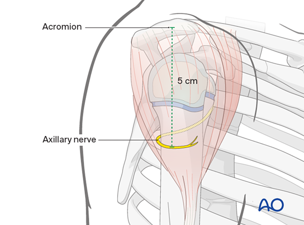
2. Axillary nerve
The most important anatomical consideration in the proximal humerus is the location of the axillary nerve, about 5 cm below the tip of the acromion in children older than 6 years.
This is particularly important in the younger child as the window for safe K-wire insertion is smaller.
The deltoid insertion is also a reproducible landmark.


